
Aberration- An optical defect. The lens does not bring all the rays of light to an exact focus. There are several different types of aberrations each having a contributing factor on image quality.
Achromatic- Color corrected optics used to produce true color.
Achromatic Condenser- A condenser corrected for spherical aberration. It is the most common type found on brightfield microscopes.
Achromatic Lens- A lens system that has been corrected to provide the same focal length for the red and blue wavelengths of light. The result is an image virtually free of extraneous coloring or aberrations.
Alignment- A condition in which all optical elements are centered on the same axis.
Analyzer- The part of a polarizing system that can select one angle of light and is used in conjunction with a polarizing filter.
Aperture- A fixed or adjustable opening or hole through which light may pass through.
Apochromat- A lens system in which chromatic aberration is corrected for three or more colors.
Apochromatic Lens- An apochromatic lens system can correct for three colors instead of two. This type of lens also has less spherical aberrations.
Arm- The part that serves as both the support of the body tube and its lens systems. The part held when the microscope is carried. Also sometimes referred to as limb.
Base- The weighted bottom portion of the microscope which gives it both balance and resistance to unexpected movement or vibration.
Beamsplitter- A device that allows light to be split into two paths usually comprised of a piece of glass with special coatings mounted at a 45 degree angle from the input source.
Binocular- A microscope head that has an eyetube for each eye.
Brightfield- An illumination method in which light is reflected off the specimen and passed through the objective to the eyepieces.
CCD-Charged Coupled Device- This is the image detector or chip inside a video camera. CCD's are categorized by their diagonal size like 1/3", 1/2", 2/3", etc.
Chromatic Aberration- An optical defect of a lens characterized as color fringes or halos. They are caused by different wavelengths of light focusing at different distances from the lens.
Coarse Adjustment- The control knob which moves the specimen (or objective) rapidly allowing focusing to occur.
Color Temperature- The quantitative value indicating the amount of color or colors emitted by an object measured in Kelvins.
Condenser- The lens system between the illuminator and the specimen which condenses the light onto the specimen.
Contrast- The ratio of light and dark. To produce a good image, you must have good contrast as well as good resolving power.
Corrected Lens- A lens or lens system which corrects for specific aberrations.
Darkfield- An illumination method used to examine specimens which cannot be distinguished from the background. Components include a dry darkfield condenser for low magnifications and any low magnification objectives. An oil darkfield condenser is used for higher magnifications. Higher magnification oil objectives must have an iris.
Daylight Filter- A blue colored filter used to correct the color temperature of a light source.
Depth of Field- A short distance along the optical axis throughout which the specimen can be seen with sufficient clarity.
Depth of Focus- A short distance along the optical axis throughout which the specimen image is focused sharply.
DIN- Deutch Industrie Normen - An international standard used in the manufacture of interchangeable objective lenses.
Disc Diaphragm- A fixed rotary disk, located under the stage of a student grade microscope, that has graded openings that allow various angles of light to come up through the stage opening. Also available as a continuously varying diameter (like a camera shutter) adjusted by a lever located on the side of the component.
Drawing Tube (Camera Lucida)- This device enables a person to view the specimen and a paper and pencil simultaneously for drawing superimposed.
Dual Viewing Attachment- Enables two people to view the same specimen through the same microscope simultaneously.
Empty Magnification- Described as magnification which increases the size, but does not increase the detail, due to the limitation of the resolving power of the optical system.
Eyepiece- The lens system closest to the eye. Also referred to as "oculars".
Eyepiece Tube- Tubes of the microscope head which hold the eyepiece.
Eye Point- The distance from the vertex of the eyepiece lens to the users eye at the eyepoint.
Eye Relief- The location or position of the eye which allows for the best possible viewing of the image.
Exit Pupil- Exit pupil describes the image that is projected to a particular point in space beyond the eyepiece. Your eye must be positioned in that exact location in order to see the full, clearly focused image. The exit pupil is a dimension usually expressed in millimeters.
Fluorescence- An illumination method used to locate fluorescently tagged material (protein, enzyme, and gene) by exciting the material with one wavelength of light in hopes that the fluorescence will appear by emitting a light at a different wavelength.
Fluorite- An objective corrected for two wavelengths and therefore, with a higher resolving power than an Achromat. There are exceptions as some manufacturers call them Fluor, Neo-Fluor, Fluotar and others.
Field- Field is the diameter of the viewing area, usually expressed in millimeters. As magnifying power is increased, the field of view is decreased.
Filter- A colored transparent material placed in the path of illumination to vary the conditions of viewing.
Filter Mount- An existing slot on the microscope which can hold filters in the path of illumination.
Filar Eyepiece- An eyepiece with an integrated measuring reticle and a moveable cross hair for making measurements on a microscope.
Fine Adjustment- The control knob which moves the specimen (or objective) very slowly allowing microtonal focusing to occur.
Finity Correction System- Finity Correction System is an optical system whereby the image formed is made solely by an objective lens.
Flatness of Field- A quality describing the appearance of the field of view as being flat from edge-to-edge.
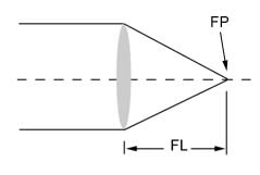
Focal Length- The distance from a point where an image is formed to a point in the lens system as shown in the drawing at right labeled "FL".
Focal Point- A point in which light rays converge after passing through a lens as shown in the drawing at right labeled "FP".
Focus Rack- The part of the microscope which allows the distance from the objective lens to the specimen to vary and to accomplish coarse and fine focus adjustments.
Huygenian Eyepiece - An eyepiece with correction for chromatic difference of magnification in the achromatic objective lens.
Illumination - The application of light onto an object or specimen under a microscope.
Illuminator - The source of light which illuminates the object or specimen to be observed. It may have fixed intensity or variable intensity via a control knob.
Incline - Eyepiece tubes are manufactured at an angle (30-45 degrees) to allow more comfortable posture during long periods of observation.
Infinity Correction System - Infinity Correction System is an optical system by which the image is formed by an objective lens and a tube lens working in tandem.
Interchangeable Eyepieces - An objective that is either threaded into a nosepiece turret or mounted on a dovetail that is removable and that can be replaced by another objective with a different magnification power.
Iris diaphragm- A device which can open and close like the iris of an eye.
JIS - Japanese Industrial Standard - An international standard used in the manufacture of interchangeable objective lenses. A Typical JIS type microscope utilizes 36mm objectives and a 170mm tube length for a 206mm system.
Koehler Illumination- Koehler illumination was introduced in 1893 by August Köhler as a method of providing the optimum specimen illumination.
Lens- An optical grade glass which has two polished surfaces and is used to converge or diverge light rays.
Light- Electromagnetic radiation consisting of photons.
Long Working Distance- Refers usually to an objective or a microscope with a greater than normal working distance.
Magnification- The enlargement of an object by an optical instrument.
Magnifying Power- The metric of a lens or combination of lenses to make an object appear larger is called magnifying power. It is the number of times the image is larger than the object would appear to the unaided eye.
Micro-manipulator- Is used to inject or extract substances from a specimen. Also used to measure or apply electric current to a specimen.
Microscope- A high precision optical instrument which uses light to observe objects. It is capable of high magnification and resolution and is used for making minute details visible.
Monocular- A microscope head with a single eyepiece.
N.A. - Numerical Aperture.- N.A is a combination of resolving power, focal depth, and luminosity of the image. The larger the N.A. value, the higher the resolving power and the smaller the focal depth.
Nosepiece- A rotary turret mounting for a set of objectives.
Objective Lens- The compound lens system in a microscope which receives light from the field of view and forms the primary image. The lens system closest to the specimen is the objective lens.
Ocular Micrometer- When an ocular micrometer is inserted in the eyepiece, it enables the person to take measurements of a specimen. It is also known as eyepiece micrometer.
Oil Immersion- Oil Immersion is the technique of placing a drop of oil between a 100X objective and the cover slip in order to improve the resolving quality of the objective. It also can be placed between the condenser and the glass slide.
Parfocal- The ability to rotate the objective turret without refocusing. With stereo microscopes, parfocal is the ability to zoom throughout the magnification range without re-focusing.
Parfocal Length- The distance between the surface of the specimen and objective lens mounting position while the specimen is in focus.
Phase Contrast- An illumination technique used to examine live, unstained specimens that have poor contrast or that are translucent.
Photomicrography- The process of documenting images on film as seen through a microscope.
Photo Port- Any port other than an eyetube which is exclusively used to connect cameras to a microscope.
Plan Objective- An objective corrected for flatness of field so that when you view the specimen it is in focus all across the image field. There are also Plan Achromats, Plan Fluorites, and Plan Apochromats.
Polarizer- A filter which can produce polarized light and is usually used in conjunction with an Analyzer.
Polarized Light- Light waves which are uniformly aligned in one direction.
Polarizing Components- Polarizing Components are components that can be added to an existing basic microscope to locate bi-refringent materials.
Prism- A solid piece of optical glass that has at least two planes inclined towards each other from which light can be reflected or refracted.
Rack and Pinion- Rack and Pinion is a mechanical design involving the intermeshing of a geared wheel and matching vertical grooved rack used in focus mechanisms.
Ramsden Eyepiece- An eyepiece similar to the Huygenian eyepiece however it has its focal plane either on or outside the surface of the collective lenses.
Reticle- A piece of glass with a pattern printed on one side that installs into an eyepiece which allows the pattern to be imposed on the microscope field of view.
Resolving Power- The ability resolve two points as two points at a given distance.
Rotational Viewing- A microscope set up in such a way that the eyepiece and eyepiece tube can rotate around horizontally. Several people gathered around the same instrument can thus view the same specimen without moving around or having to move the microscope.
Spherical Aberration- A spherical aberration is an optical defect by which the lens fails to form a sharp image. Rays of light which pass through a lens near its edge are converged to a point nearer the lens than those rays passing through the center of the lens.
Stage- The platform which holds the specimen. Types include: plain, mechanical, motorized, heated, etc.
Mechanical Stage- Moves the specimen East to West and North to South via X-Y controls.
Rotating Stage - Can usually rotate 360 degrees or, in the case of a rotating mechanical stage, rotate as much as 270 degrees.
Stage Clips- Chrome metal Fasteners located on the stage and placed over slides to hold them securely in place while viewing.
Teaching Head- An accessory that allows more than one user to see into the microscope simultaneously.
Trinocular- A microscope head with two eyepieces and a photo port.
Tube Length- The Tube Length is the optical distance from the objective to the eyepiece. Tube Length governs the interchangeability of optical components; i.e. a microscope objective corrected for 160mm tube length cannot be used on a microscope corrected for infinity.
Turret-mounted - Objectives that are attached to one common rotating nosepiece that allows for quick and accurate objective positioning during viewing.
Ultra-violet- The portion of the spectrum of light where the wavelengths are above the visible spectrum.
Video Microscope- Enables a specimen to be viewed on a video screen. Image can also be analyzed by a computer using image analysis software.
Virtual Image- Virtual Image is an image that does not converge in open space.
Wavelength- Light travels in waves varying in length. The measurement of a light wave is from the top of one wave to the top of the next one and is usually measured in units of nanometers (nm) or Angstroms (A).
Wide Field Eyepiece- A Wide Field Eyepiece is an eyepiece having a large field of view with a high eye point.
Working Distance- The distance from the objective to the specimen with the image in focus.


