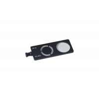
MT-50/BFDF- SLIDER- Darkfield / Brightfield Slider for 4X-40X objectives . Slides into standard BF Condenser slot of MT-50 and MT-51.
Inserts the dark-field into condenser socket and pull the dark-field diaphragm in then it can fulfill simply dark-field observation. You can observe blood , flagellum, Treponema palliduim in Darkfield at any times between 4X -40X with this accessory easily.
Light microscopy is an important investigative tool for biology that is used regularly in high schools and colleges. The structure of many biological specimens are of low contrast that cannot be revealed by the brightfield compound microscopes which are provided in many classrooms. Microscopes that improve the contrast of these specimens through special optics are prohibitively expensive for most teaching budgets. The MT-50/BFDF- SLIDER is a simple, inexpensive modification that changes a brightfield microscope into a darkfield microscope allowing low contrast samples to be examined. This explains the basic theory of darkfield microscopy. This technique allows expanded use of the brightfield microscope at all levels of teaching with very little manipulation or expense.
Details
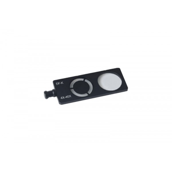
MT-50/BFDF- SLIDER- Darkfield / Brightfield Slider for 4X-40X objectives . Slides into standard BF Condenser slot of MT-50 and MT-51.
Theory of darkfield microscopy
Microscopes are used to magnify objects.
Through magnification, an image is made to appear larger than the original object. The magnification of an object can be calculated roughly by multiplying the magnification of the objective lens times the magnification of the ocular lens (eyepieces).
Objects are magnified to be able to see small details. There is no limit to the magnification that can be achieved; however, there is a magnification beyond which detail does not become clearer. The result is called empty magnification when objects are made bigger but their details do not become clearer.
Therefore, not only magnification but resolution is important to the quality of the of an image.
The resolving power of the microscope is defined as the ability to distinguish two points apart from each other. The resolution of a microscope is dependent on a number of factors in its construction. There is also an inherent theoretical limit to resolution imposed by the wavelength of visible light (400-600nm). The theoretical limit of resolution (the smallest distance able to be seen between two points) is calculated as:
Resolution = 0.61?/N.A.
Where ? represents the wavelength of light used and N.A.is the numerical aperture. The student-grade microscopes generally have much lower resolution than the theoretical limit because of lower quality lenses and illumination systems.
Standard brightfield microscopy relies upon light from the lamp source being gathered by the substage condenser and shaped into a cone whose apex is focused at the plane of the specimen. Specimens are seen because of their ability to change the speed and the path of the light passing through them. This ability is dependent upon the refractive index and the opacity of the specimen. To see a specimen in a brightfield microscope, the light rays passing through it must be changed sufficiently to be able to interfere with each other which produces contrast (differences in light intensities) and, thereby, build an image. If the specimen has a refractive index too similar to the surrounding medium between the microscope stage and the objective lens, it will not be seen. To visualize biological materials well, the materials must have this inherent contrast caused by the proper refractive indices or be artificially stained. These limitations require instructors to find naturally high contrast materials or to enhance contrast by staining them which often requires killing them. Adequately visualizing transparent living materials or thin unstained specimens is not possible with a brightfield microscope.
Darkfield microscopy relies on a different illumination system. Rather than illuminating the sample with a filled cone of light, the condenser is designed to form a hollow cone of light. The light at the apex of the cone is focused at the plane of the specimen; as this light moves past the specimen plane it spreads again into a hollow cone. The objective lens sits in the dark hollow of this cone; although the light travels around and past the objective lens, no rays enter it The entire field appears dark when there is no sample on the microscope stage; thus the name darkfield microscopy. When a sample is on the stage, the light at the apex of the cone strikes it. The image is made only by those rays scattered by the sample and captured in the objective lens The image appears bright against the dark background. This situation can be compared to the glittery appearance of dust particles in a dark room illuminated by strong shafts of light coming in through a side window. The dust particles are very small, but are easily seen when they scatter the light rays. This is the working principle of darkfield microscopy and explains how the image of low contrast material is created: an object will be seen against a dark background if it scatters light which is captured with the proper device such as an objective lens.
The highest quality darkfield microscopes are equipped with specialized costly condensers constructed only for darkfield application. This darkfield effect can be achieved in a brightfield microscope, however, by the addition of a simple "stop". The stop is a piece of opaque material placed below the substage condenser; it blocks out the center of the beam of light coming from the base of the microscope and forms the hollow cone of light needed for darkfield illumination.
If a manufactured darkfield stop is not available for your microscopes, there are some alternatives. If there is a filter holder below the condenser, a dark field stop from another company may fit or be made to fit. This alternative can be a bit tricky because the material needs to be placed in the center of the condenser. Technically, to properly block the beam, the stop should vary in diameter from 8mm to 20mm depending on the magnification and numerical aperture of the objective lens.
Darkfield microscopy reduces the amount of light entering the lens system of a microscope in two ways. First, the stop blocks the center of the beam of light that would otherwise fill the objective lens. Second, only the light which is scattered by the specimen and enters the objective lens is seen. Therefore, the best viewing result requires increasing the light intensity as much as possible: by setting the light intensity adjustment at maximum, by opening the field diaphragm, by opening the condenser aperture, and by removing any color or other filters. The correct microscope slides also should be used; they should be 1mm thick.
The illumination needs to be aligned and adjusted to achieve the best image. Before making the darkfield modification, align the light beam in the center of the field of view according to the manufacturer¹s instructions. To facilitate focusing the substage condenser and the objective lens, use a slide filled with samples that are easy to find; instructions for making a cheek cell slide follow. Focus the sample slide at low magnification (10X) in the brightfield mode. Insert the darkfield stop without changing the focus. Make sure that the maximum amount of light is available. Rack up the condenser to its highest position with the condenser focus knob. Look at the sample and slowly lower the condenser until the sample is visible against a dark background and in sharpest contrast. Finally, adjust the view of the image with the fine focus knob.
Limitation and Advantages of darkfield microscopy
The advantage of darkfield microscopy also becomes its disadvantage: not only the specimen, but dust and other particles scatter the light and are easily observed. For example, not only the cheek cells but the bacteria in saliva are evident. More care in sample preparation needs to be exercised in darkfield applications. Glass slides need to be thoroughly cleaned of extraneous dust and dirt. It may be necessary to filter sample media (agar, water, saline) to exclude confusing contaminants. Sample materials need to be spread thinly; too much material on the slide creates many overlapping layers and edges making it difficult to interpret structures.
To create the image, this technique relies on scattered light from specimens. Color is lacking or minimal; this can be disappointing to the viewer. The actual size of specimens is also impacted; the width of objects becomes exaggerated.
Examples of darkfield images.
Living Chlamydomonas, a biflagellate algae in brightfield and in darkfield modes. Although the cells are seen with brightfield, the flagella are not discernible . Some contrast can be achieved by closing down the condenser aperture diaphragm which then allows the flagella to be seen However, closing down the condenser aperture decreases the numerical aperture of objective lens effectively reducing the resolution. Darkfield allows flagella and details inside the to be seen distinctly The use of darkfield microscopy thus achieves both high contrast and high resolution.
Cheek cells make a quick study. Buccal cells are obtained by gently scraping the inside of the mouth with a toothpick and thinly spreading the cells on a slide; a cover slip is placed over the preparation which is not allowed to dry. These cells have no inherent contrast and are difficult to see with a brightfield. Dramatic contrast is achieved in a darkfield microscope; the nucleus and other intracellular inclusions as well as bacteria in the surrounding medium can be easily located and identified.