|
Model
|
Head
|
Illumination
|
Eyepieces
|
Objectives
|
Stage
|
Condenser
|
|
TC-5100 Brightfield |
Binocular
|
Koehler w/o iris LED illumination
|
SWH10x FN22
|
Quintuple Nosepiece w/LWD 10X &
LWD 20X |
Plain Fixed Stage
180mm X 247mm |
TC Condenser
WD 73mm NA 0.30orTC Condenser2 WD 20.5mm NA 0.55 |
|
TC-5100E Brightfield |
Ergonomic Binocular | |||||
|
TC-5200 Brightfield |
Trinocular
|
|||||
|
TC-5300 and TC-5300L Phase Contrast |
Binocular |
Koehler w/o iris
with phase slider LED transmitted |
||||
|
TC-5300E Phase Contrast |
Ergonomic Binocular
|
|||||
|
TC-5400 and TC-5400L Phase Contrast |
Trinocular | |||||
|
TC-5500 Epi-Fluorescent |
Binocular | 100W Mercury + Koehler w/o iris LED Transmitted |
Quintuple Nosepiece |
TC Condenser WD 73mm |
||
|
TC-5600 Epi-Fluorescent |
Trinocular |
Includes:
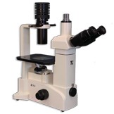
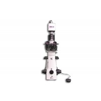
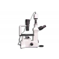
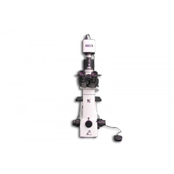
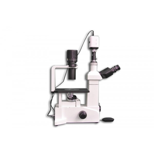
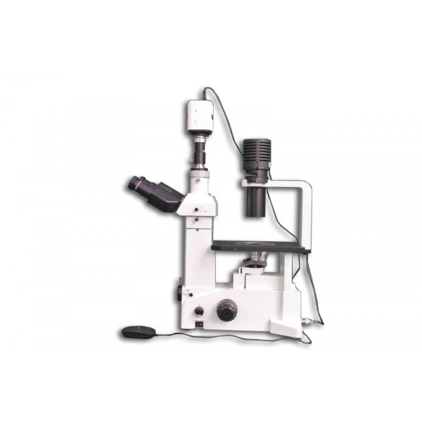
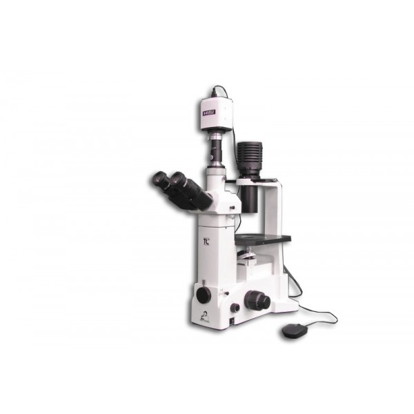
• TC-5400L: Trinocular Inverted Phase Contrast/Brightfield Compound microscope, with ∞Infinity Corrected Optical System, F-200MM with LED Koehler illumination with quintuple nosepiece and auto voltage sensing power supply
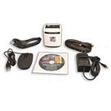
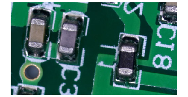
• HD1500T: The HD1500T is our full featured 6 MP camera with a 1/3” CMOS chip.The HD1500T features a full HD (1080x1920) resolution, which provides high fidelity images with a refresh rate of 60fps- frames per second when connected via HDMI, so there is absolutely no lag in the live video feed when moving your specimen or part.
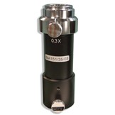
• MA151/35/03: C-mount adapter with 0.3X lens
• MA816/10: Siedentopf type trinocular head, inclined at 30°, Interpupillary adjustment 53mm to 75mm graduated diopter on left eyetube (30mm I.D. eyetubes)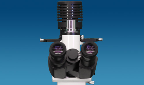
• MA817 (x 2): SWH10X Widefield high eyepoint eyepiece, Field No. 22, tube O.D. 30.0 mm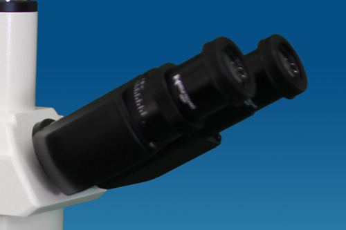
• MA826: TC Long Working Distance LWD Planachromat Phase 10X objective ∞/1.0
• MA827: TC Long Working Distance LWD Planachromat Phase 20X objective ∞/1.0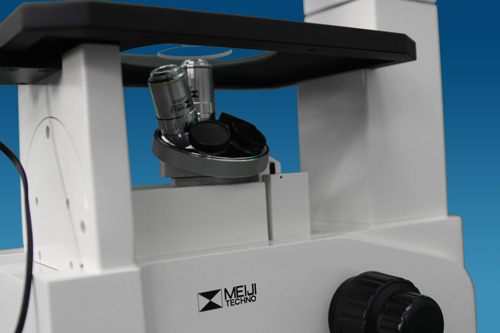
• MA853: TC Extra Long Working Distance ELWD Condenser, N.A. 0.30, W.D. 73.0mm
• MA861: Green interference filter in 51mm mount (546mm)
• MA863: Cobalt clear filter in mount in 51mm mount
• STAGE: Plain stage with 180mm(X) x 245mm(Y) plain stage with replaceable glass insert with 45mm opening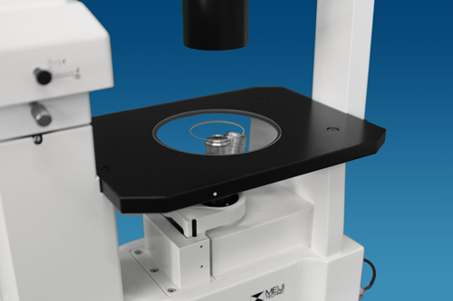
• MA878: Glass Stage plate insert for plain stage (Round-109mm diameter, 1.5mm thick with 44.3mm central opening)
• MA855: Phase slider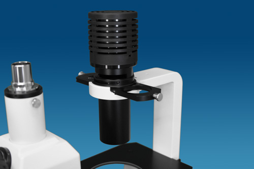
• MA458/05: Centering telescope (O.D. = 30.0)
• MA809/10: Removable AC Electric cord with plug for 115V models
• MA326: Bulb LED (Factory Installed)
• MA327: Fuse 3 Amp (Factory Installed)
• Manual: TC-5000 Meiji Techno Instruction Manual
• Warranty: Limited Lifetime Warranty Card
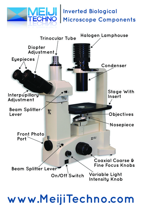
VIEWING HEADS
Binocular models, the TC5100, TC5300 and TC5500, incorporate the MA814 Siedentopf-type binocular head while the trinocular models, the TC5200, TC5400 and TC5600 have the MA816/10 Siedentopf-type trinocular head. The MA816/10 uses an 80/20 beamsplitter that can be engaged for photo-microscopy. (100% to eyetubes or 80% to phototube and 20% to the eyetube.) Each microscope head has the eyetubes inclined at 30 degrees with the left eyetube having graduated diopter settings. The interpupillary distance on the viewing heads is adjustable between 53mm - 75mm.
EYEPIECES
10X Super Widefield High Eyepoint eyepieces are standard and 15X and 20X eyepieces are also available as an option. A Super Widefield High Eyepoint 10X focusable eyepiece that accepts standard 25mm reticules is also available.
OBJECTIVE CHANGER
Quintuple nosepiece provides effortless objective changes with smooth-operating, ball bearing mounted.
OBJECTIVES
Meiji Techno America offers an assortment of Planachromat Infinity Corrected (ICOS) objectives for Brightfield, Darkfield and Phase Contrast observation modes.
STAGE
The optional attachable mechanical stage measures 112mm(X) x 72mm(Y). It has ergonomically positioned right-handed coaxial drop down controls. It also accepts the following optional accessory plates: 35mm, 55mm, 65mm, Petri Dish Holders, 96-well Terasaki Plate Holder, 1" X 3" glass slide holder, Hemacytometer holder.Slide, chamber, and petri dish holding plates of various sizes are available separately.
CONDENSER
The standard condenser has a working distance of 73mm.
ILLUMINATION
A powerful LED transmitted with pre-centered socket provides enhanced image quality and brightness for the observation of specimens.
PHOTOMICROGRAPHY
Meiji Techno offers a broad range of digital video cameras which offers excellent sensitivity, high resolution and a wide dynamic range for the most demanding brightfield and darkfield microscopy applications including clinical pathology and cytology, life sciences and geology. Some models has included software, that can preview and capture still shots and live videos to a PC or Mac with USB port or HDMI connectivity.
HD1500T Info:
• HD1500T Camera
• HDMI Cable
• USB Cable
• 12V AC/DC Power adapter
• SD Card
• Mouse
• Application Software ISCapture
• Packing Box
Package Information:
• 5lbs
• 12 x 10 x 4 inches
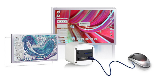
NEW: BUILT-IN MOUSE CONTROL CAMERA
The significant innovation of the HD15000T is making the software implant inside the camera. This forward thinking feature frees users from cumbersone computers and annoying buttons. You can control the camera by only a mouse directly.
Videos of the camera in action with our microscopes:
Brief overview with the compare feature
With Darkfield microscopy.
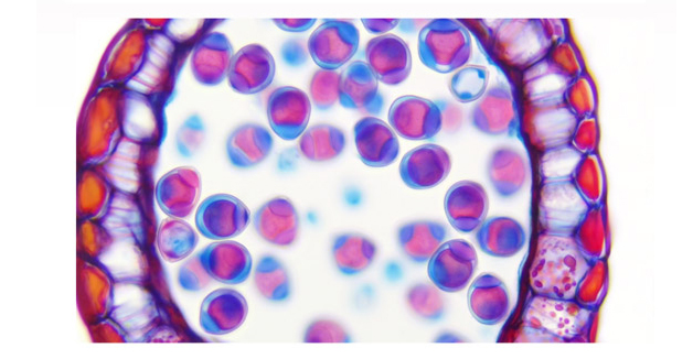
PERFECT COLOR REPRODUCTION
Meiji Techno's HD technology offers true-to-life reproduction oh image color. What you see on the monitor is what you get from the microscope eyepiece. No compressed date transmission, resulting in scientific-grade images.
1080P 60FPS FULL FRAME RATES
With 60fps at 1080p, the HD1500T presents a perfect combination of outstanding resolution and frame rate. This allows the user to fluently manipulate the live images without any lags.
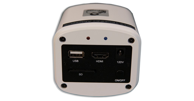
HDMI+USB 2.0+SD OUTPUT
Multiple output options are available for this powerful HDMI camera. HDMI 1080P, USB 2.0, or SD memory card can be used individually or all three SIMULTANEOUSLY.
DUAL FPGA PROCESSORS
With an innovative Dual FPGA processor architecture design, FPGA1 for image quality processing and FPGA2 for image output control, you get a high-speed preview without sacrificing image quality. It is the most powerful camera you deserve to have.
SMART PRESENTATION
Camera parameters such as exposure time, white balance, and gain are designed by default to operate automatically. Automatic optimum configuration is expected to be done in 0.015 seconds, and you'll find that the camera is easily managed from the start.
ADVANCED PARAMETER SETTING
For users with specific requirements, the camera is designed with parameter setting buttons that enable manual adjustment of white balance, gain, saturation, Gamma and screen freeze.
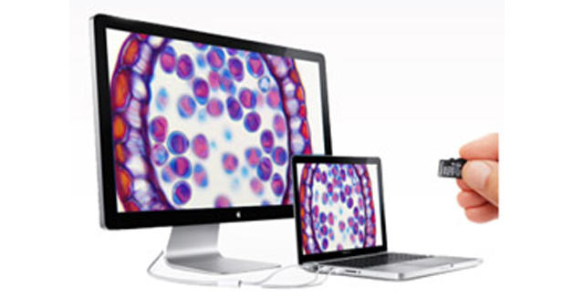
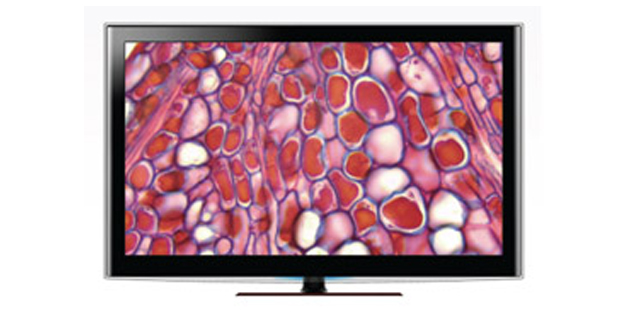
PC TO TV
Images are displayable on PCs and HDMI-installed TVs. All parameters can be controlled by the computer to adjust them to actual situations. This is advantageous in teaching and meeting applications.
On-board Video Saving
The SD card can save not only still images, but also dynamic images in 720P color definition, such that motion trails are recorded in fast speed and intact form.
LOW LIGHT SENSITIVITY
Thanks to its highly sensitive sensor, the HD1500T is capable of capturing low-light images rapidly. In darkfield imaging and even fluorescence, it will automatically increase the sensitivity, presenting low light images with clarity in high-speed preview mode.
|
Model
|
Type
|
Version
|
Mega Pixels
|
Resolution
|
Frame Rate (FPS)
|
Sensor
|
C" Mount
|
|
HD1500T |
HDMI/USB 2.0 | Color | 2-6MP | 3264 x 1840 Static 1920 x 1080 Dynamic |
60fps by HDMI 30fps by USB 2.0 |
1/2.8" CMOS | 0.3X |
|
HD1500TM |
HDMI/USB 2.0 | Color | 2-6MP |
3264 x 1840 Static |
60fps by HDMI 30fps by USB 2.0 |
1/2.8" CMOS | 0.3X |
|
HD1500MET |
HDMI/USB 2.0 | Color | 2-6MP | 3264 x 1840 Static 1920 x 1080 Dynamic |
60fps by HDMI 30fps by USB 2.0 |
1/2.8" CMOS | 0.3X |
|
HD1500MET-M |
HDMI/USB 2.0 | Color | 2-6MP |
3264 x 1840 Static |
60fps by HDMI 30fps by USB 2.0 |
1/2.8" CMOS | 0.3X |
|
HD1500MET-AF |
HDMI/USB2.0 | Color | 2-6MP |
3264 x 1840 Static |
60fps by HDMI 30fps by USB 2.0 |
1/2.8" CMOS | 0.3X |
|
HD1500MET-M-AF |
HDMI/USB2.0 | Color | 2-6MP |
3264 x 1840 Static |
60fps by HDMI 30fps by USB 2.0 |
1/2.8" CMOS | 0.3X |
|
HD1000-LITE |
HDMI/USB 2.0 | Color | 5MP |
2592 x 1944 |
15fps by HDMI |
1/2.5" CMOS |
0.3X |
|
HD1000-LITE-M |
HDMI/USB 2.0 | Color | 5MP |
2592 x 1944 |
15fps by HDMI |
1/2.5" CMOS |
0.3X |
|
HD1600T |
USB 3.0 | Color | 16MP | 4608 x 3456 |
25fps by USB 3.0 |
1/2.33" CMOS |
0.3X |
|
SS500-MC |
Wifi | Color | 5MP | 2912 x 1640 | 60fps by Wifi | 1/2.3" CMOS | 0.3X |
|
WF300 |
Wifi | Color | 8MP | 3200 x 2400 | 60fps by USB 2.0 30fps by Wifi |
1/2.8" CMOS | 20-43mm Eyepiece Mount |
|
WF200 |
Wifi | Color | 5MP | 2592 x 1944 | 40fps by USB 2.0 15fps by Wifi |
1/2.8" CMOS | 0.3X |
|
WF100 |
Wifi | Color | 5MP | 2592 x 1944 | 30fps by Wifi | - | - |
|
X-1000U |
USB 2.0 | Color | 5MP | 2592 x 1911 | 10fps by USB 2.0 | 1/2.8" CMOS | 0.3X |
|
MIS-PL-DV24 |
USB 2.0 | Color | 2MP | 1600 x 1200 | 30fps by USB 2.0 | 1/2" CMOS | 0.5X |
|
DK-LITE-B |
USB 2.0 | Color | 1.5MP | 1440 x 1080 | 10fps by USB 2.0 | 1/2.5" CMOS | 0.5X |
|
DK1000CB |
USB 2.0 | Color | 2MP | 1600 x 1200 | 15fps by USB 2.0 | 1/2" CMOS | 0.5X |
|
DK1000M |
USB 2.0 | Monochrome | 1.3MP | 1280 x 1024 | 30fps by USB 2.0 | 1/2" CMOS | 0.5X |
|
DK3000C |
USB 2.0 | Color | 3.1MP | 2048 x 1536 | 12fps by USB 2.0 | 1/2" CMOS | 0.5X |
|
DK5000C |
USB 2.0 | Color | 5MP | 2592 x 1944 | 5fps by USB 2.0 | 1/2.5" CMOS | 0.7X |
|
HD2000C |
USB 2.0 | Color | 2MP | 1920 x 1080 | 60fps by USB 2.0 | 1/3" CMOS | 0.3X |
|
ST1000C |
USB 3.0 | Color | 5MP | 2592 x 1944 | 60fps by USB 3.0 | 1/2.5" CMOS | 0.5X |
Product Information:
• Manufactured: 100% MADE IN JAPAN
• Warranty: LIMITED LIFETIME WARRANTY
• Customer Support: 1-(800) 832-0060 (U.S.A Based)
TC-5400L Package Information:
• Weight: ~35 lbs (15.8 kg)
• Box Size: 19 x 15 x 25 inches
• Trinocular head type: 485 mm(D) x 440 mm(H) x 2210 mm(W) (8.5Kg)(31.0 lbs)
HD1500T Package Information:
• 3lbs
• 12 x 10 x 4 inches
The TC-5000 Brightfield/Phase Contrast Series features include:
Brightfield:
The simplest of all the optical microscopy illumination techniques. Sample illumination is transmitted (i.e., illuminated from below and observed from above) white light and contrast in the sample is caused by absorbance of some of the transmitted light in dense areas of the sample. Brightfield microscopy is the simplest of a range of techniques used for illumination of samples in light microscopes and its simplicity makes it a popular technique. The typical appearance of a brightfield microscopy image is a dark sample on a bright background.
Phase Contrast:
An optical microscopy technique that converts phase shifts in light passing through a transparent specimen to brightness changes in the image. Phase shifts themselves are invisible, but become visible when shown as brightness variations. When light waves travel through a medium other than vacuum, interaction with the medium causes the wave amplitude and phase to change in a manner dependent on properties of the medium. Changes in amplitude (brightness) arise from the scattering and absorption of light, which is often wavelength-dependent and may give rise to colors. Photographic equipment and the human eye are only sensitive to amplitude variations. Without special arrangements, phase changes are therefore invisible.
The TC-5300 & TC-5400 Series are suitable for viewing colorless and transparent specimens and live cells. The TC-5300 & TC-5400 Series is a contrast-enhancing optical technique that can be utilized to produce high-contrast images of transparent specimens such as living cells, microorganisms, thin tissue slices, lithographic patterns, and sub-cellular particles (such as nuclei and other organelles). In effect, the phase contrast technique employs an optical mechanism to translate minute variations in phase into corresponding changes in amplitude, which can be visualized as differences in image contrast. One of the major advantages of phase contrast microscopy is that living cells can be examined in their natural state without being killed, fixed, and stained. As a result, the dynamics of ongoing biological processes in live cells can be observed and recorded in high contrast with sharp clarity of minute specimen detail.


|
Model
|
Head
|
Illumination
|
Eyepieces
|
Objectives
|
Stage
|
Condenser
|
|
TC-5100 Brightfield |
Binocular
|
Koehler w/o iris LED illumination
|
SWH10x FN22
|
Quintuple Nosepiece w/LWD 10X &
LWD 20X |
Plain Fixed Stage
180mm X 247mm |
TC Condenser
WD 73mm NA 0.30orTC Condenser2 WD 20.5mm NA 0.55 |
|
TC-5100E Brightfield |
Ergonomic Binocular | |||||
|
TC-5200 Brightfield |
Trinocular
|
|||||
|
TC-5300 and TC-5300L Phase Contrast |
Binocular |
Koehler w/o iris
with phase slider LED transmitted |
||||
|
TC-5300E Phase Contrast |
Ergonomic Binocular
|
|||||
|
TC-5400 and TC-5400L Phase Contrast |
Trinocular | |||||
|
TC-5500 Epi-Fluorescent |
Binocular | 100W Mercury + Koehler w/o iris LED Transmitted |
Quintuple Nosepiece |
TC Condenser WD 73mm |
||
|
TC-5600 Epi-Fluorescent |
Trinocular |
Included:
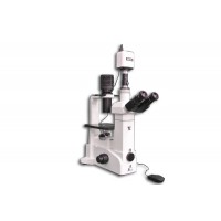
• TC-5400: Trinocular Inverted Phase Contrast/Brightfield Compound microscope, with ∞Infinity Corrected Optical System, F-200MM with LED illumination with quintuple nosepiece and auto voltage sensing power supply
• MA816/10: Siedentopf type trinocular head, inclined at 30°, Interpupillary adjustment 53mm to 75mm graduated diopter on left eyetube (30mm I.D. eyetubes)
• MA817 (x 2): SWH10X Widefield high eyepoint eyepiece, Field No. 22, tube O.D. 30.0 mm
• MA826: TC Long Working Distance LWD Planachromat Phase 10X objective ∞/1.0
• MA827: TC Long Working Distance LWD Planachromat Phase 20X objective ∞/1.0
• MA853: TC Extra Long Working Distance ELWD Condenser, N.A. 0.30, W.D. 73.0mm
• MA861: Green interference filter in 51mm mount (546mm)
• MA863: Cobalt clear filter in mount in 51mm mount
• STAGE: Plain stage with 180mm(X) x 245mm(Y) plain stage with replaceable glass insert with 45mm opening
• MA878: Glass Stage plate insert for plain stage (Round-109mm diameter, 1.5mm thick with 44.3mm central opening)
• MA855: Phase slider
• MA458/05: Centering telescope (O.D. = 30.0)
• MA809/10: Removable AC Electric cord with plug for 115V models
• MA326: Bulb 6V 30W Halogen (Factory Installed)
• MA327: Fuse 3 Amp (Factory Installed)
• HD1500T: The HD1500T is our full featured 6 MP camera with a 1/3” CMOS chip.The HD1500T features a full HD (1080x1920) resolution, which provides high fidelity images with a refresh rate of 60fps- frames per second when connected via HDMI, so there is absolutely no lag in the live video feed when moving your specimen or part.
• MA151/35/03: C-mount adapter with 0.3X lens
OPTIONAL COMPONENTS & ACCESSORIES BELOW
Viewing Heads:
• MA814: Siedentopf type binocular, inclined at 30°, Interpupillary adjustment 53mm to 75mm graduated diopter on left eyetube (30mm I.D. eyetubes)
• MA957/15: Ergonomic binocular head, inclination adjustable vertically from 10° to 50° (Included with TC-5100E)
• MA1034: Trinocular Siedentopf head, erect image, inclined at 30°, I.P. adjustment 53mm to 75mm, graduated diopter on left eyetube (30mm I.D. eyetubes) Includes 0/100 beamsplitter (100% to eyetubes or 100% to phototube)
Eyepieces:
• MA818: SWH 15X Widefield high eyepoint eyepiece, Field No. 16.0, tube O.D. 30.0 mm (each) (19mm reticle mount)
• MA819: SWH 20X Widefield high eyepoint eyepiece, Field No. 12.0, tube O.D. 30.0 mm (each) (19mm reticle mount)
• MA859: SWH 10X-F Focusing eyepiece with reticle mount (without reticle), tube O.D. 30.0 mm (each) (25mm reticle mount)
Phase Objectives:
• MA825: TC Planachromat Phase 4X objective ∞/1.0 N.A. 0.13, F.L. 50.0, W.D. 17.3
• MA828: TC Planachromat Phase 40X objective ∞/1.0 N.A. 0.65, F.L. 5.0, W.D. 2.8
Condensers:
• MA854: TC Condenser, N.A. 0.55, W.D. 20.5mm (For use with MA823 40X objective)
Filters:
• MA856: LB100 Blue clear filter in 51mm mount
• MA857: G533 Green clear filter in 51mm mount
• MA858: ND25 Neutral density filter in 51mm mount
Miscellaneous:
• MA809/20: Removable AC Electric cord with plug for 220V models (included in all 220V models) CAMERA ADAPTERS FOR “C” MOUNT TYPE CAMERAS
• MA877: “C” mount type camera adapter for CCD/CMOS camera (No internal lens) (Magnification factor of 0.66X from TC5000 Series front camera port)
CAMERA ADAPTERS FOR DIGITAL STILL CAMERAS
• MA868: Digital camera adapter for non “C” mount type cameras (52mm O.D. thread) For TC Series front camera port only: (Requires additional DCA adapter ring for your specific model of camera which are listed on the last two pages of the TC5000 section) CAMERA ADAPTERS FOR 35mm TYPE CAMERAS
• MA869: 35mm SLR camera attachment Magnification at film plane is 2.23X of objective in use
Stages (Fixed 245mm x 180mm, plain stage is included with stand):
• MA878: Replacement glass stage plate insert for plain stage - TC5000 Series (Round - 109mm diameter, 1.5mm thick with 44.3mm opening) (Fits plain stage only not for use with MA380/05)
• MA380/05: Attachable mechanical stage, movement 112mm(X) x 72mm(Y) with test plate holder and drop-down coaxial X-Y controls.
COMPONENTS OFTEN PAIRED WITH THE MA380/05 MECHANICAL STAGE
Specimen Holders for use with MA380/05 Attachable Mechanical Stage (Rectangular Plate 129.74mm x 89.78 mm x 3mm):
• MA382: Slide glass holder, 1”x 3”
• MA383: Chamber holder (For 55mm x 80mm chamber)
• MA384: Petri dish holder 35mm diameter
• MA385: Petri dish holder 55mm diameter
• MA387: Petri dish holder 65mm diameter
• MA864: Terasaki plate holder (For 56mm x 87mm Terasaki plate)
• MA378: Hemacytometer holder
Stage Micrometers:
• MA285: Objective micrometer, 0.01mm ruling
• MA286: 0.04” divided into 40 units, 0.001
• MA285: 1mm divided into 100 parts
Eyepiece Micrometers:
• MA523: Cross-line reticule, 25mm diameter
• MA506: Eyepiece micrometer, 10mm divided into 100 units, 25mm diameter
• MA509: Eyepiece micrometer 5mm divided into 100 units, 25mm diameter
• MA524: 10mm square divided into 100 parts, 1.0mm square, 25mm diameter
• MA542: Cross-line with 0.1mm graduations, 21mm diameter
Double click to read at full screen
Download Brochure (PDF)
Double click to read at full screen
Download Manual (PDF)
Download Camera Driver (ZIP)
Download TCapture Software (ZIP)
Download MAC ISListen Capture Software (2018)
Download ISListen MAC Installation Guide (PDF)
Double click to read at full screen
Download Manual (PDF)
Double click to read at full screen
Download Software Manual (PDF)
Double click to read at full screen
Download Menu Quick Guide (PDF)
Double click to read at full screen
Download Calibrate Guide (PDF)
Double click to read at full screen
Download FAQ Sheet (PDF)
Double click to read at full screen
Download Manual (PDF)