Meiji Techno is the third-largest manufacturer of optical microscopes in Japan.
Originally a manufacturer of educational microscopes, Meiji Techno has since extended product lines into the industrial, laboratory, and higher education markets. Our products are sold throughout Europe, Asia, Africa and the Americas by agents or distributors authorized by Meiji Techno Co., Ltd.
During the years Meiji Techno has opened a representative offices in different parts of the world (America, Great Britain, Europe, Russia) and has established a wide dealer’s network that united high-quality specialists having experience and knowledge in various scientific and technical areas.
Meiji Techno confirms high quality of the products by offering 5-years guarantee. Meiji Techno uses high-quality own-produced optical and mechanical components for production of microscopes. These products can operate for many years in case of providing proper maintenance and following operation guidelines. Microscopes having easy and reliable construction can operate in hard conditions of industrial enterprises. The quality of Meiji Techno microscopes is approved by international certificates.
Convert your Meiji microscope into a digital scanner!
Specifications:
| Technical Specifications | |
| Software |
DSS Basic |
| Camera | |
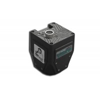
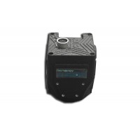
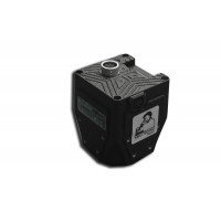
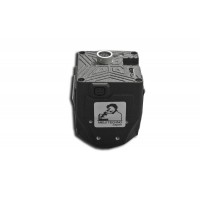
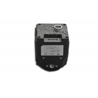
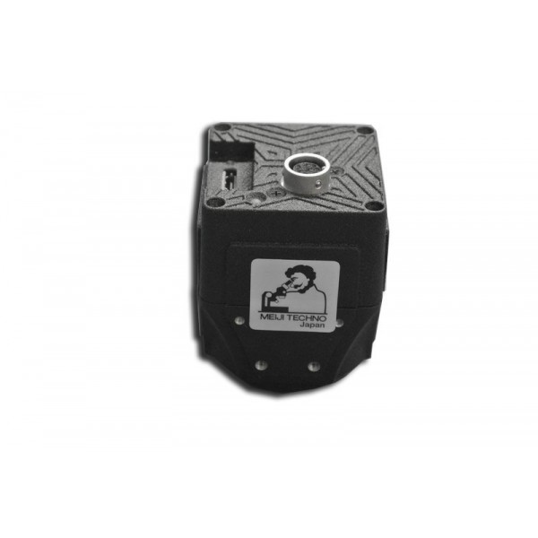
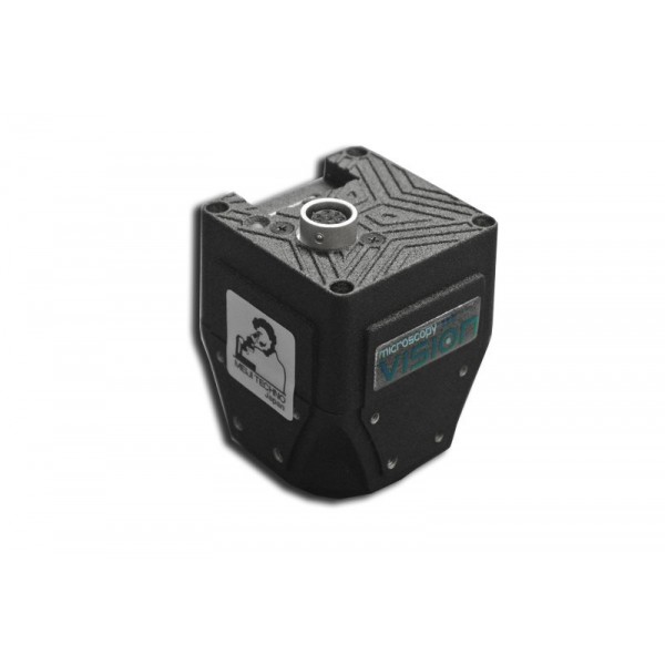
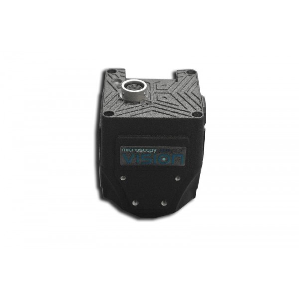
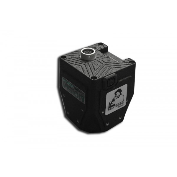
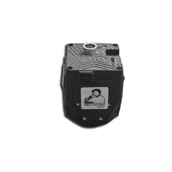
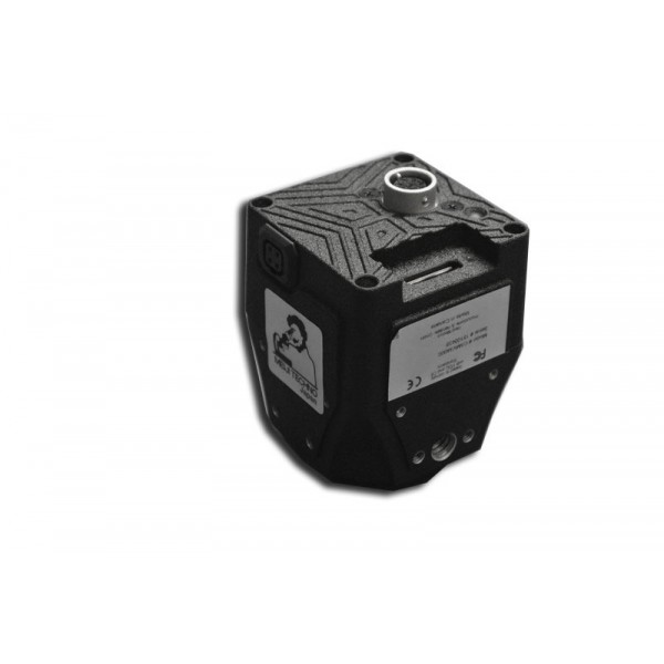
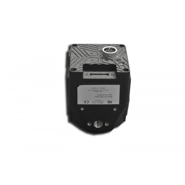
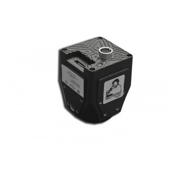
| Vision CAM 3200 | 2.8 MP, resolution 2064 x 1544, image sensor CMOS 1/1.8", ~120 f/s, USB 3.0 |
| PC | |
| DSS Basic system requirements |
- Intel Core i5 processor |
| Microscope | |
| Requirements | Trinocular Meiji Techno microscope with Plan Achromat objectives (or better) |
| Imagine modes* | Brightfield, dark field, phase contrast and oil immersion |
| Application fields | Digital pathology and microscopy, reaserch and education, biology and medicine |
DSS Basic Initial set includes:
DSS Basic software, digital camera Vision CAM® V3200 - Use your PC and monitor
Meiji Techno / Saitama, Japan: to create panoramic microscopy images with high resolution it is often required to use expensive scanning devices. Meiji Techno has developed DSS Basic – a solution, combining software for image processing and camera, that allows to perform scanning using a common biological microscope. DSS Basic will be introduced first at ASCB international forum (American Society of Cell Biology) that will take place in San-Diego, the USA, on December 8th-12th, 2018.
In clinical pathology and scientific research, conclusions are often based on microscopy images of tissues and other results of investigation. For the purposes of documentation or independent opinion, a digital high-resolution panoramic image of the tested sample is extremely valuable. However, the corresponding part of the information consists of separate cells, or the morphological area of interest is much smaller than the complete sample. Thus, at present time special scanners are used for scanning a complete sample; but these solutions are not available for majority of specialists. DSS Basic is a solution for scanning that can be easily integrated with a microscope and is available for pricing.
Meiji Techno has developed an easy and convenient alternative to automatic slide scanners. DSS Basic is a solution to create wide-scale scanning using manual microscope and camera. Automatic way to proceed the images stitches separate microscopy images in real time - without motorized microscope needed. During the stitching process, the images are automatically placed to create a panoramic view that is known as virtual slide. Integrated additional functions make DSS Basic an effective and easy solution for exchanging data.
High-quality imaging data speed up diagnostics
DSS Basic creates panoramic images – scanning large squares – in real time mode, that allows to speed up diagnostics and document results. DSS Basic is easy to use and extremely versatile instrument, that can be integrated in existing laboratory information systems. DSS Basic can be installed on any microscope.
Application areas in clinical diagnostics and material science
DSS Basic can contribute to various application areas, like telemedicine, documentation systems and education of doctors and biologists. Application is also possible in other fields where high-resolution microscopy images are playing an important role. Various scientific research in biology and medicine, quality control and testing different materials can be referred to them.
Easy digital scanning and full visualization of slides
Efficient solution for scanning in microscopy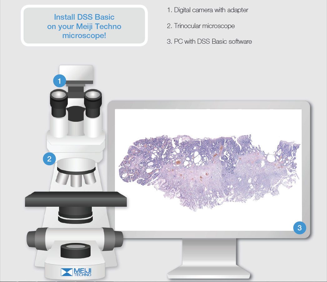
How does it work?
• Install the camera on the microscope and the software on your PC
• Press start and move the sample
• Digital slide forms automatically as you move the sample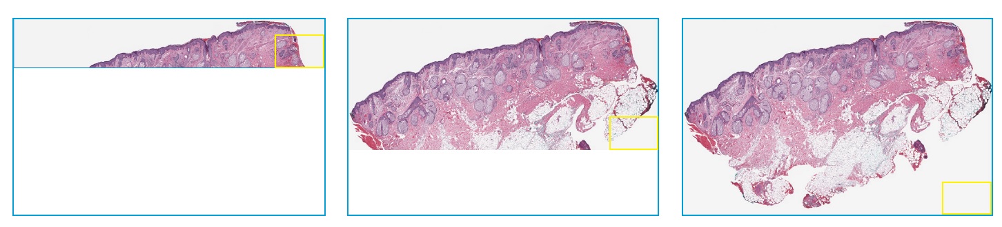
Digital Slide Analysis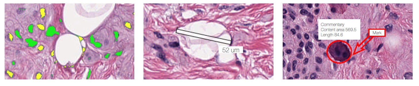
Object analysis
Objects classification by specified criteria
Count and classification
Automatic or manual object identification on the digital slide. Measurements of linear and optical parameteres for particles.
Marks and comments
Make your comments and put marks on a digital slide


Double click to read at full screen
Download Press Release (PDF)
Double click to read at full screen
Download Brochure (PDF)


