|
Model
|
Head
|
Illumination
|
Eyepieces
|
Objectives
|
Stage
|
Condenser
|
|
MT4210L |
Binocular
|
LED Illumination
|
KHW10x FN20
|
Plan 4X,
S.Plan Ph10x, S.Plan Ph40x |
Fixed flat-top left or right-handed controls
|
Turret Type
NA 1.25 |
|
MT4210EL |
Ergonomic Binocular | |||||
|
MT4310L |
Trinocular
|
|||||
|
MT4310EL |
Ergonomic Trinocular | |||||
|
MT4210H |
Binocular |
6V 30W halogen
|
||||
|
MT4210EH |
Ergonomic Binocular
|
|||||
|
MT4310H |
Trinocular | |||||
|
MT4310EH |
Ergonomic Trinocular
|
Includes:
• MT4310EL: Ergonomic Trinocular Phase Contrast/Brightfield Compound microscope with Bright LED Koehler illumination with quintuple nosepiece and auto voltage sensing power supply
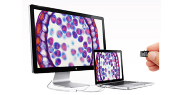
• HD1500TM: The HD1500TM’s display features full HD (1080x1920) resolution and provides high fidelity images from the HD1500TM with a refresh rate of 60 frames per second. Included with the display is a mounting kit that allows the display to be mounted directly on the HD1500TM.
• MA151/35/03: C-mount adapter with 0.3X lens
• MA957/05: Ergonomic binocular head, inclination adjustable from 10-50 degrees
• MA958: Photo/video attachment with sliding 80/20 beam-splitter. For use with video or digital cameras when MA957/05 ergonomic head is used
• MA407 (x 2): KHW10X compensating eyepiece, Field No. 20, Tube O.D. 23.2MM (each)
• MA831: U. Plan Objective 4X, N.A 0.10, W.D. 25.8mm, F.N. 24
• MA839: U. Plan Phase Objective 10X, N.A. 0.25, W.D. 10.7mm, F.N. 24
• MA841: U. Plan Phase Objective 40X, N.A. 0.65, W.D. 0.5mm, F.N. 24
• MA458: Centering Telescope, 23.2mm OD
• MA913: Zernike Phase Condenser with centerable phase annuli for 10X, 20X, 40X, 100X with brightfield position, dark- field stop for 40X objective
• MA857/05: Green Filter, Clear, 44.0mm Diameter, unmounted
• MA917R: Ceramic coated flat-top stage, 170mm x 140mm, 80mm x 52mm
• MA598: Attachable finger assembly for MA917R and MA917L
• LED/SP-1: 3W LED, 5500K color temperature (Factory Installed)
• MA929: Spare fuse 0.5A for MT4310EL LED Illuminator (Factory Installed)
• MA809/10: Removable AC Electric cord with plug for 115V models
• MA701: Dust Cover for MT4310EL
• Manual: MT4310EL Meiji Techno Instruction Manual
• Warranty: Limited Lifetime Warranty Card
Product Information:
• Manufactured: 100% MADE IN JAPAN
• Warranty: LIMITED LIFETIME WARRANTY
• Customer Support: 1-(800) 832-0060 (U.S.A Based)
Package Information:
• Weight: 29 lbs (8.0kg)
• Box Size: 16 x 14 x 21 inches
• Footprint: 263mm (D) x 176mm (W)
• Binocular head type: 390 mm(D) x 410mm (H) x 210mm(W) (8.0kg)( 29.0 lbs)
• Trinocular head type: 390 mm(D) x 465mm(H) x 210mm(W) (8.7Kg)( 30.0 lbs)
• Binocular Ergonomic head type: 390 mm(D) x 483mm (H) x 210mm(W) (8.4kg)(31 lbs)
• Trinocular Ergonomic head type: 390 mm(D) x 533mm(H) x 210mm(W) (8.9Kg)(33 lbs)
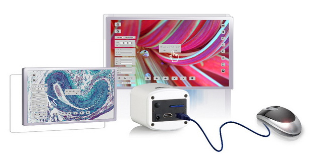
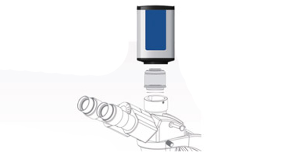
HD1500MET-M: The HD1500MET-M is our full featured 6 MP camera with a 1/3” CMOS chip. A 0.3X C” mount adapter (MA151/35/03) is needed to attach it to any standard Meiji Techno America Trinocular Microscope. The HD1500MET-M features a full HD (1080x1920) resolution, which provides high fidelity images with a refresh rate of 60fps- frames per second when connected via HDMI, so there is absolutely no lag in the live video feed when moving your specimen or part. Included with the 11.8” High Definition display is a mounting kit that allows the display to be mounted directly in front of the HD1500MET-M.
• MA151/35/03: C-mount adapter with 0.3X lens
• MA879/05: CHROMA filter-AT-GFP/FITC longpass-19002 mounted in Meiji Filter cube, Exciter(EX) AT480/30x, Dichroic (BS)AT505DC, Emitter(EM)AT5151p (Factory Installed)
• MA880/05: CHROMA filter-AT-TRITC/Cy3 Longpass-19004 mounted Meiji filter cube, Exciter (EX)AT540/25x, Dichroic (BS)AT565DC, Emitter(EM)575lp (Factory Installed)
• MA881/05: CHROMA filter-AT-DAPI/Hoechst/AlexaFlour 350-3900 mounted in Meiji filter cube, Exciter (EX):AT375/28x, Dichroic (BS):AT415DC, Emitter (EM):AT460/50m (Factory Installed)
• FLPWJ: D.C. Power Supply (This power supply is for the 100W Mercury lamp only)
• MA860: ND25 Neutral Density filter, 29.8mm diameter
• MT-RF47: UV Eye protection filter
• PE-300L/LEDMT: LED fluorescence illumination Kit, configured for fluorophores such a DAPI,FITC,TRITC & Cy5, Instant on/off with the ability to optimize intensity and minimize sample damage via the simple desktop Control Pod with a manual control pod and powe supply (Included in MT6200CL & MT6200ECL & MT6300CL & MT6300ECL models)
PE-300L/LEDMT features:
- Direct fit to the microscope in seconds
- Broad spectrun - covers UV (DAPI) to Red region (Cy5)
- No alignment, a once only adjustment
- Instant on/off - no shutters required, no warm up or cool down
- Stable and repeatable - reliable and precise results
- Fine intensity control in 1% steps (0-100%) - no ND filters required
- Excellent uniformity over field of view - fixed and stable, no alignment necessary
• MA816: Trinocular head-Siedentopf type, I.P. adjustment 53mm to 75mm, graduated diopter on left eyetube (30.0mm I.D. eyetube). Inclined at 30 degrees, Inverted image
• MA817 (x 2): SWH10X Widefield high eyepoint eyepiece, Field No. 22, tube O.D. 30.0 mm
• MA844: Plan Semi Apo Objective F10X, N.A.0.40, W.D.1.0 mm, F.N.24
• MA845: Plan Semi Apo Objective F20X, N.A. O.65, W.D. 0.7 mm, F.N.24
• MA846: Plan Semi Apo Objective F40X, N.A. 0.82, W.D. 0.15 mm, F.N.24
• MA848: Plan Semi Apo Objective F100X oil, N.A. 1.25, W.D.0.2mm, F.N.24
• MA700: Immersion oil
• MA910: Abbe condenser, N.A.1.25 with iris diaphragm and dove tail mount
• MA856/05: LB100 Blue Clear filter, 44.0mm diameter, unmounted
• MA918R: Ceramic coated flat-top stage, 190mm x 149mm, with 80mm x 52mm X-Y movement, with right hand drop down coaxial controls
• MA598: Attachable finger assembly for MA918R and MA918L
• MA326: 6V 30W Halogen lamp (Factory Installed)
• MA327: 3 Amp Fuse for Halogen illuminator
• MA809/10: Removable AC Electric cord with plug (115V)
• MA906 (x 2): Rubber Eyeshield for MA817 SWH10X eyepiece (Factory installed)
• MA714: Dust Cover for MT6000 Series
• Manual: MT6000 Meiji Techno Instruction Manual
• Warranty: Limited Lifetime Warranty Card
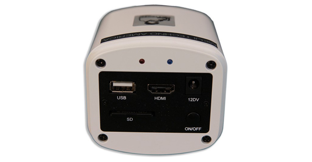
HD1500TM Includes:
• HD1500TM Camera
• HDMI Cable
• USB Cable
• 12V AC/DC Power adapter
• SD Card
• Mouse
• Application Software ISCapture
• Packing Box
Package Information:
• 5lbs
• 12 x 10 x 4 inches
NEW: BUILT-IN MOUSE CONTROL CAMERA
The significant innovation of the HD15000T is making the software implant inside the camera. This forward thinking feature frees users from cumbersone computers and annoying buttons. You can control the camera by only a mouse directly.
Videos of the camera in action with our microscopes:
Brief overview with the compare feature
With Darkfield microscopy.
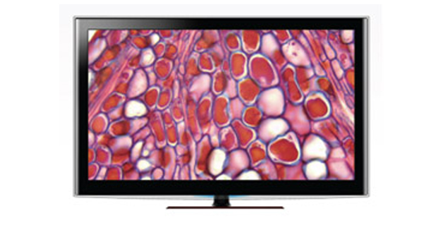
PERFECT COLOR REPRODUCTION
Meiji Techno's HD technology offers true-to-life reproduction oh image color. What you see on the monitor is what you get from the microscope eyepiece. No compressed date transmission, resulting in scientific-grade images.
1080P 60FPS FULL FRAME RATES
With 60fps at 1080p, the HD1500T presents a perfect combination of outstanding resolution and frame rate. This allows the user to fluently manipulate the live images without any lags.
HDMI+USB 2.0+SD OUTPUT
Multiple output options are available for this powerful HDMI camera. HDMI 1080P, USB 2.0, or SD memory card can be used individually or all three SIMULTANEOUSLY.
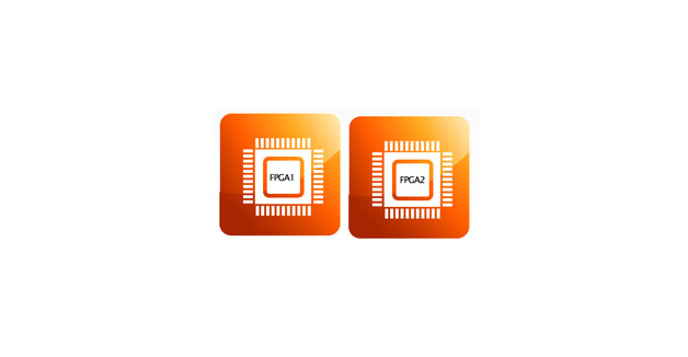
DUAL FPGA PROCESSORS
With an innovative Dual FPGA processor architecture design, FPGA1 for image quality processing and FPGA2 for image output control, you get a high-speed preview without sacrificing image quality. It is the most powerful camera you deserve to have.
SMART PRESENTATION
Camera parameters such as exposure time, white balance, and gain are designed by default to operate automatically. Automatic optimum configuration is expected to be done in 0.015 seconds, and you'll find that the camera is easily managed from the start.
ADVANCED PARAMETER SETTING
For users with specific requirements, the camera is designed with parameter setting buttons that enable manual adjustment of white balance, gain, saturation, Gamma and screen freeze.
PC TO TV
Images are displayable on PCs and HDMI-installed TVs. All parameters can be controlled by the computer to adjust them to actual situations. This is advantageous in teaching and meeting applications.
On-board Video Saving
The SD card can save not only still images, but also dynamic images in 720P color definition, such that motion trails are recorded in fast speed and intact form.
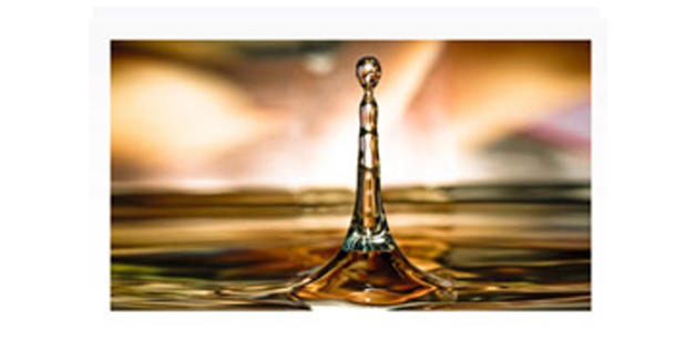
LOW LIGHT SENSITIVITY
Thanks to its highly sensitive sensor, the HD1500T is capable of capturing low-light images rapidly. In darkfield imaging and even fluorescence, it will automatically increase the sensitivity, presenting low light images with clarity in high-speed preview mode.
|
Model
|
Type
|
Version
|
Mega Pixels
|
Resolution
|
Frame Rate (FPS)
|
Sensor
|
C" Mount
|
|
HD1500T |
HDMI/USB 2.0 | Color | 2-6MP | 3264 x 1840 Static 1920 x 1080 Dynamic |
60fps by HDMI 30fps by USB 2.0 |
1/2.8" CMOS | 0.3X |
|
HD1500TM |
HDMI/USB 2.0 | Color | 2-6MP |
3264 x 1840 Static |
60fps by HDMI 30fps by USB 2.0 |
1/2.8" CMOS | 0.3X |
|
HD1500MET |
HDMI/USB 2.0 | Color | 2-6MP | 3264 x 1840 Static 1920 x 1080 Dynamic |
60fps by HDMI 30fps by USB 2.0 |
1/2.8" CMOS | 0.3X |
|
HD1500MET-M |
HDMI/USB 2.0 | Color | 2-6MP |
3264 x 1840 Static |
60fps by HDMI 30fps by USB 2.0 |
1/2.8" CMOS | 0.3X |
|
HD1500MET-AF |
HDMI/USB2.0 | Color | 2-6MP |
3264 x 1840 Static |
60fps by HDMI 30fps by USB 2.0 |
1/2.8" CMOS | 0.3X |
|
HD1500MET-M-AF |
HDMI/USB2.0 | Color | 2-6MP |
3264 x 1840 Static |
60fps by HDMI 30fps by USB 2.0 |
1/2.8" CMOS | 0.3X |
|
HD1000-LITE |
HDMI/USB 2.0 | Color | 5MP |
2592 x 1944 |
15fps by HDMI |
1/2.5" CMOS |
0.3X |
|
HD1000-LITE-M |
HDMI/USB 2.0 | Color | 5MP |
2592 x 1944 |
15fps by HDMI |
1/2.5" CMOS |
0.3X |
|
HD1600T |
USB 3.0 | Color | 16MP | 4608 x 3456 |
25fps by USB 3.0 |
1/2.33" CMOS |
0.3X |
|
SS500-MC |
Wifi | Color | 5MP | 2912 x 1640 | 60fps by Wifi | 1/2.3" CMOS | 0.3X |
|
WF300 |
Wifi | Color | 8MP | 3200 x 2400 | 60fps by USB 2.0 30fps by Wifi |
1/2.8" CMOS | 20-43mm Eyepiece Mount |
|
WF200 |
Wifi | Color | 5MP | 2592 x 1944 | 40fps by USB 2.0 15fps by Wifi |
1/2.8" CMOS | 0.3X |
|
WF100 |
Wifi | Color | 5MP | 2592 x 1944 | 30fps by Wifi | - | - |
|
X-1000U |
USB 2.0 | Color | 5MP | 2592 x 1911 | 10fps by USB 2.0 | 1/2.8" CMOS | 0.3X |
|
MIS-PL-DV24 |
USB 2.0 | Color | 2MP | 1600 x 1200 | 30fps by USB 2.0 | 1/2" CMOS | 0.5X |
|
DK-LITE-B |
USB 2.0 | Color | 1.5MP | 1440 x 1080 | 10fps by USB 2.0 | 1/2.5" CMOS | 0.5X |
|
DK1000CB |
USB 2.0 | Color | 2MP | 1600 x 1200 | 15fps by USB 2.0 | 1/2" CMOS | 0.5X |
|
DK1000M |
USB 2.0 | Monochrome | 1.3MP | 1280 x 1024 | 30fps by USB 2.0 | 1/2" CMOS | 0.5X |
|
DK3000C |
USB 2.0 | Color | 3.1MP | 2048 x 1536 | 12fps by USB 2.0 | 1/2" CMOS | 0.5X |
|
DK5000C |
USB 2.0 | Color | 5MP | 2592 x 1944 | 5fps by USB 2.0 | 1/2.5" CMOS | 0.7X |
|
HD2000C |
USB 2.0 | Color | 2MP | 1920 x 1080 | 60fps by USB 2.0 | 1/3" CMOS | 0.3X |
|
ST1000C |
USB 3.0 | Color | 5MP | 2592 x 1944 | 60fps by USB 3.0 | 1/2.5" CMOS | 0.5X |
Frequently Asked Questions
Q: Start the software and get the camera recognized, but the preview is all black.
A: Check whether the anti-dust cap on the C-mount is removed. If already removed and attach the camera to the microscop, check whether the lighting path toggle switch to the camera. Then try to extend the exposure time and gain to get brighter images.
Q: The imaging scene from the eyepiece is in focus but the live images from the camera are still out of focus.
A: This is because the camera and the eyepiece are in different focal planes. Can consider to use adjustable adapter to connect camera to the microscope trinocular, and the adjust the adapter to make the live image in focus.
Q: There are some shadows on the image and the shadows always stay in the same position even when the sample is moved.
A: Possible reasons:
1. The anti-dust film on the camera C-mount is not removed. Remove the anti-dust film
2. Check to see if there is any dirt on the objective. Clean the objective.
3. Some dust on the surface of the camera IR filter. Clean the camera optical port.
Q: Camera frame rate is extremely slow.
A: Possible reasons:
1. Preview resolution is too high. Try lowering the resolution and check the change of the frame rate.
2. Exposure time is too long. If need exposure time longer than 200ms, try to use higher gain to get brighter images.
3. Computer issue. Computer is very old or the motherboard chipset is Intel 8 Series/C220 series.
Q: Start the software and get the error message: can not recognize the camera or No camera.
A: Possible reasons:
1. The camera USB cable is not well connected. Reconnect the camera again.
2. Check whether the camera is connect to the USB port on the front of the mainframe. If so, then connect it to the USB port on the back of the mainframe. The USB ports on the front of the mainframe are the extended ports from the motherboard which can not provide stable voltage and current for cameras.
3. The camera itself needs extra power supply but does not switch on the power. TrueChrome is required to switch on power, attach to the PC and then restart the software. TCC-6.1ICE is required to plug in power to be recognized by PC.
4. The camera driver is not installed or failed to install. Go to the "Device Manager" > "Imaging Devices" and check whether the camera is listed WITH YELLOW FLAG. Then re-install the driver again. If still fail to install to driver, please contact camera technical support for help.
Q: Image Field of View (FOV) is much smaller than the scene can be seen from the eyepieces.
A: Possible reasons:
The camera sensor size is not 1 inch. It needs reduce lens to get bigger FOV.
Low resolution is used to preview. Set full resolutionion.
The reduce lens does not match the camera sensor size.
Q: When push “Capture” button, sometimes the saved images are quite blurry.
A: It could be because some slight vibration happens on the camera during capturing the images.
Q: Security shield on ISCapture shortcut.
A: Some anti-virus or firewall softwaree installed in the PCs will identify the DLL files from ISCapture as virus and automatically isolate or delete the ISCapture software files. If have the warning message to delete or isolate the DLL files during the ISCapture installation, please ignore the warning message or set the ISCapture as trusted software. Otherwise, the ISCapture can not be used normally.
Q: Newer version software con not recognize the old cameras.
A: Currently, all the newer version software ISCapture is compatible with the previous version cameras EXCEPT TCA Series.
Q: After doing the measurement on the live images, the captured image is without any measurement data.
A: To save the live measurement on captured images, check the “Combined Measurements” checkbox in the File Save tab and then capture images. While doing the still image measurements, only click the Save or Save As on the right hand side shortcut to save the data on the images.
Q: The live images and the captures images color is quite different from the one we can see from the eyepieces.
A: Because of the different lighting source, White Balance is needed to correct the image color.
Q: How to do quick White Balance?
A: White Balance while attached to camera to stereo microscope: Use a white paper to replace the sample. Click on “White Balance” to correct the image color > Remove the white paper and put back the sample.
White Balance while attach the camera to the biological microscope: Move the sample slide to the blank area. Click on “White Balance” and move back the sample.
Q: After install ISCapture and firstly start the software, but get the error “The program can’t start because MSVCP100.dll is missing from your computer. Try reinstalling the program to fix this problem.”

A; This is because the operating system lack of some Visual C++ 32bit library files which the ISCapture needs to call them. Without these files, the software can not be started normally. Install the patch VCredist_x86.exe (can be found in the CD that comes with the camera or go to http://www.microsoft.com/en-us/download/details.aspx?id=5555
Q: Start the ISCapture but get the error “Unable to execute file C:\Program Files\ISCapture\ISCapture.exe CreateProcess failed; code 740.
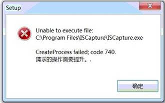
A: Completely remove the ISC Capture, reboot the PC and then right click on ISCapture installer (ISCapture Setup.exe) and select “Run as Administrator” to install the ISCapture. When you first start the software right click on the ISC icon and select “Run as Administrator” to start it.
Q:When start ISCapture, get error message “Cannot find the USB Key!”

A: The software is encrypted. Specific USB key is needed to attach to the computer port first.
Q:After capturing several images, it takes more and more time to save a frame image. Sometimes it is even up to more than 30 seconds, and then finally the software hang or crash.
A: If the image save directory is the network driver, during uploading the files to network driver, it will significantly increase the COP usage. When the CUP usage is up to 100%, the software will hang or crash. Recommend to change the file save directory to the computer hard disk.
Q: Whether can share the calibration files on different PCs?
A: If using the ISCapture earlier version than V4.0: Go to the folder “Application Data/ISCapture” in system disk, find the file “calibrationTable.ini” and copy it to another PC under the same directory. (The “Application Data” folder is a hidden folder, you might need to make the system invisible visible first.)
If it is ISCapture V4.0 or later, go to the Parameter tab and click “Backup” to backup the parameter settings and copy them to another PC to restore them.
Q:After install the ISCapture and every time I run the software i get the error:
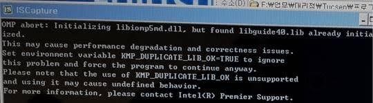
A: This is because some program in the user’s PC is using the library file ‘libiomp5md.dll’which will conflict with the ISCapture. So far as we know, the software GoPro Studio will conflict with ISCapture. To solve this issue, please try to remove the libiomp5md.dll from the C:\Windows\System32 folder while using the ISCapture.
Q: Shortcut keys for ISCapture to capture still images and videos:
A: With ISCapture V3.6.6 or later, running the ISCapture first, Press F9 key on the keyboard to capture images. Press F10 to start the video recording. If in manual recording mode, press F11 to stop the recording.
Q: On preview images, lots of horizontal lines appear.

A: This is because of the lighting frequency. Extend exposure time or turn off the filament lamp or fluorescent lamp in the environment to solve this issue.
Q:Recording issue when connected to the HD1500T/HD1000 Series to the monitor and recorded video directly to the SD card? If so, do you know what the capacity of the SD card the user used during recording the videos?
A:When recording videos to the SD card, the camera only supports the SD card with format SDHC. It can not work with the SD card format SDXC. And max. capacity of the SDHC format SD card is 32GB which means the SD card with capacity >= 64GB, is format SDXC.
If already use SDHC format SD card, please help to confirm the information below:
1). Whether the customer used the SD card came with the HD1500T/HD1000 camera?
2). If not, what exactly the model of the SD card the customer used? Maybe some picture of the SD card.
If got the issue while connect the camera to the computer, I need to confirm below information:
1). The software always stop recording AUTOMATICALLY after 5-10 minutes?
2). Whether the software or the computer freeze if MANUALLY stop the recording?
Product Information:
• Manufactured: 100% MADE IN JAPAN / 100% MADE IN UK (CoolLED)
• Warranty: LIMITED LIFETIME WARRANTY
• Customer Support: 1-(800) 832-0060 (U.S.A Based)
MT4310EL Package Information:
• Weight: 29 lbs (8.0kg)
• Box Size: 16 x 14 x 21 inches
• Footprint: 263mm (D) x 176mm (W)
• Trinocular Ergonomic head type: 390 mm(D) x 533mm(H) x 210mm(W) (8.9Kg)(33 lbs)
HD1500TM Package Information:
• 5lbs
• 12 x 10 x 4 inches
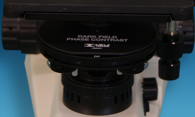
Meiji Techno Phase Contrast optical components needs to be aligned to the condenser annulus with the phase plate in the objective rear focal plane with a phase telescope, that inserts into one of the microscope eyepiece observation ports. The phase contrast annulus is used in the substage condenser on Meiji Techno’s upright microscope within the condenser turret. The Phase ring in the condenser must be specifically matched to the particular Phase objective equipped with a corresponding phase plate annulus. For example, the Phase 10X and Phase 20X both of which contain a phase plate will require a condenser annuli having different diameters that correspond to the objective magnification and N.A. (Numerical Aperture). By properly matching the Meiji Techno Condenser annulus to the objective phase plate, the Meiji Techno Phase microscope can be aligned to superimpose illuminating light rays passed through the annulus onto the Phase ring of the objective to achieve proper Phase contrast illumination.
The Ph1 condenser annulus , which contains the smallest aperture, is designed to be used with the lower power 10X and 20X Phase objective. Intermediate magnification objective 40X Phase utilize the Ph 2 annulus, while the highest power and N.A. 100X objective require the Ph 3 annulus, which requires the largest phase annulus.
The Ph1 position in the Phase Condenser Turret facilitates Objectives Phase 10X and Phase 20X
The Ph2 position in the Phase Condenser Turret facilitates Objectives Phase 40X
The Ph3 position in the Phase Condenser Turret facilitates Objectives Phase 100X
The MT4000 Phase Microscopes features include:
Brightfield:
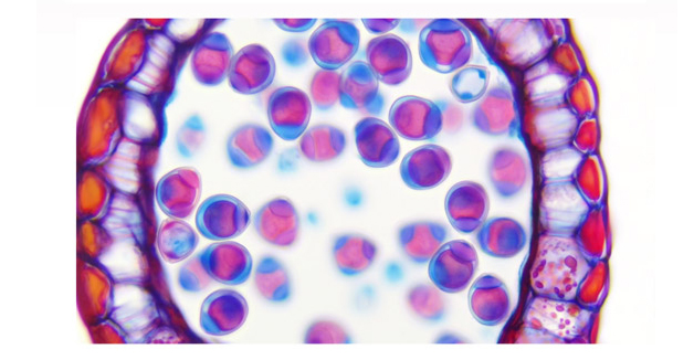
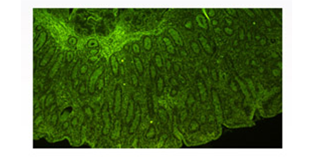
The simplest of all the optical microscopy illumination techniques. Sample illumination is transmitted (i.e., illuminated from below and observed from above) white light and contrast in the sample is caused by absorbance of some of the transmitted light in dense areas of the sample. Brightfield microscopy is the simplest of a range of techniques used for illumination of samples in light microscopes and its simplicity makes it a popular technique. The typical appearance of a brightfield microscopy image is a dark sample on a bright background.
Phase Contrast:
An optical microscopy technique that converts phase shifts in light passing through a transparent specimen to brightness changes in the image. Phase shifts themselves are invisible, but become visible when shown as brightness variations. When light waves travel through a medium other than vacuum, interaction with the medium causes the wave amplitude and phase to change in a manner dependent on properties of the medium. Changes in amplitude (brightness) arise from the scattering and absorption of light, which is often wavelength-dependent and may give rise to colors. Photographic equipment and the human eye are only sensitive to amplitude variations. Without special arrangements, phase changes are therefore invisible.
The MT4000 Series is a newly designed upright biological compound microscope system. Ergonomically designed, precisely manufactured and simple to use stage and coarse and fine controls. To meet the demands of researchers and clinical laboratory specialists. The MT4000 Series was to develop to provide functionality and operational ease. Viewing high quality images, sample changing and photomicroscopy are all conducted with ergonomics in mind. High luminescent LED illumination reduces need for frequent bulb replacement but still provide rugged durability and long life span.
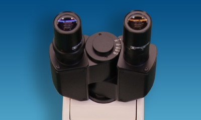
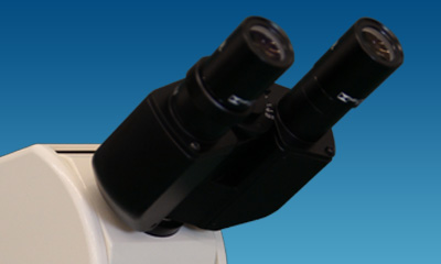
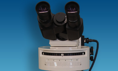
VIEWING HEADS
Model MA815/05 is the Siedentopf-type binocular head and Model MA816/05 is the trinocular head for camera integration. Each head has the eyetubes inclined at 30 ° with the left eyetube having graduated diopter settings. The interpupillary distance is adjustable between 53mm - 75mm. An 80/20 beamsplitter for the trinocular tube can be engaged for photo work.The optional MA957/05, Ergonomic Binocular Viewing Head, has the inclination adjustable from 10 to 50 degrees to fit users of different heights. The Ergonomic Trinocular model comes with the MA958, photo/video attachment with sliding 80/20 beam-splitter (Included in MT4300ED).
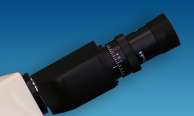
EYEPIECES
10X Widefield High Eyepoint eyepieces F.N.20 are standard, and 15X and 20X eyepieces are available as an option. A Widefield High Eyepoint 10X focusable eyepiece that accepts 21mm reticules is also available.
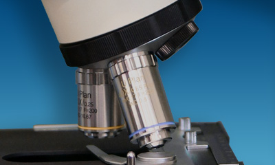
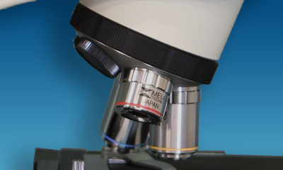
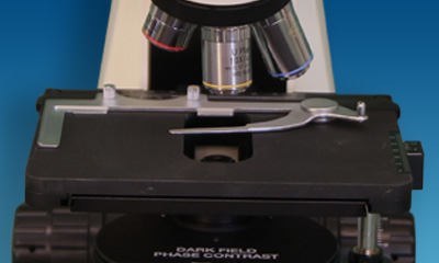
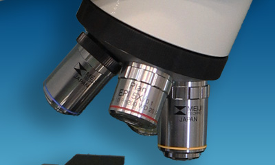
OBJECTIVE CHANGER
Quintuple nosepiece provides effortless objective changes with smooth-operating, ball bearing mounted.
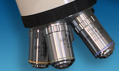
OBJECTIVES
Meiji Techno America offers an assortment of Planachromat Infinity Corrected (ICOS) objectives for Brightfield, Darkfield and Phase Contrast observation modes.
STAGE
Ceramic coated standard right-handed or optional left-handed controls, flat top stage 171mm x 115mm and travels 78mm(X) x 52mm(Y). Ergonomically positioned coaxial drop down controls. Available with motorized focus and stage controls.
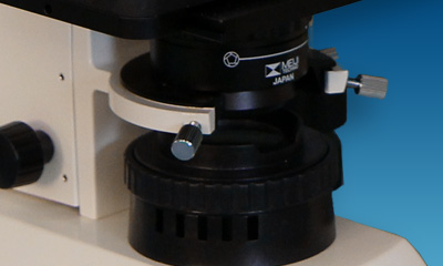
CONDENSER
New design centerable Abbe 1.25 N.A. Condenser with built-in iris diaphragm in dovetail mount.
ILLUMINATION
Powerful white LED illumination provides enhanced image quality and brightness for the observation of specimens and photomicroscopy.
PHOTOMICROGRAPHY
Meiji Techno offers a broad range of digital video cameras which offers excellent sensitivity, high resolution and a wide dynamic range for the most demanding brightfield and darkfield microscopy applications including clinical pathology and cytology, life sciences and geology. Some models has included software, that can preview and capture still shots and live videos to a PC or Mac with USB port or HDMI connectivity.
 Model
Model|
Model
|
Head
|
Illumination
|
Eyepieces
|
Objectives
|
Stage
|
Condenser
|
|
MT4210L |
Binocular
|
LED Illumination
|
KHW10x FN20
|
Plan 4X,
S.Plan Ph10x, S.Plan Ph40x |
Fixed flat-top left or right-handed controls
|
Turret Type
NA 1.25 |
|
MT4210EL |
Ergonomic Binocular | |||||
|
MT4310L |
Trinocular
|
|||||
|
MT4310EL |
Ergonomic Trinocular | |||||
|
MT4210H |
Binocular |
6V 30W halogen
|
||||
|
MT4210EH |
Ergonomic Binocular
|
|||||
|
MT4310H |
Trinocular | |||||
|
MT4310EL |
Ergonomic Trinocular
|
Included:
• MT4310EL: Ergonomic Trinocular Phase Contrast/Brightfield Compound microscope with Bright LED Koehler illumination with quintuple nosepiece and auto voltage sensing power supply
• MA957/05: Ergonomic binocular head, inclination adjustable from 10-50 degrees
• MA958: Photo/video attachment with sliding 80/20 beam-splitter. For use with video or digital cameras when MA957/05 ergonomic head is used
• MA407 (x 2): KHW10X compensating eyepiece, Field No. 20, Tube O.D. 23.2MM (each)
• MA831: U. Plan Objective 4X, N.A 0.10, W.D. 25.8mm, F.N. 24
• MA839: U. Plan Phase Objective 10X, N.A. 0.25, W.D. 10.7mm, F.N. 24
• MA841: U. Plan Phase Objective 40X, N.A. 0.65, W.D. 0.5mm, F.N. 24
• MA910: Abbe condenser, N.A. 1.25 with iris diaphragm and dove-tail mount
• MA917R: Ceramic coated flat-top stage, 170mm x 140mm, 80mm x 52mm
• MA598: Attachable finger assembly for MA917R and MA917L
• LED/SP-1: 3W LED, 5500K color temperature
• MA929: Spare fuse 0.5A for MT4000 LED Illuminator
• MA809/10: Removable AC Electric cord with plug for 115V models
• MA701: Dust Cover for MT4000
• HD1500TM: The HD1500TM’s display features full HD (1080x1920) resolution and provides high fidelity images from the HD1500TM with a refresh rate of 60 frames per second. Included with the display is a mounting kit that allows the display to be mounted directly on the HD1500TM.
• MA151/35/03: C-mount adapter with 0.3X lens
OPTIONAL COMPONENTS & ACCESSORIES BELOW
Viewing Heads:
• MA816/05: Trinocular head - Siedentopf type, I.P. adjustment 53mm to 75mm, graduated diopter on left eyetube (23.2mm I.D. eyetube). Inclined at 30 degrees (Included in Model MT4300L, MT4300H, MT4310L and Model MT4310H) Inverted image
• MA957/05: Ergonomic binocular head, inclination adjustable from 10-50 degrees (23.2mm I.D. eyetube) Erect image. (Included in all Ergonomic Bino. & Trino. models)
Photo/Video Attachment:
• MA958: Photo / video attachment with sliding 80/20 beam-splitter. For use with video or digital cameras when MA957/05 ergonomic head is used (Included in all Ergonomic Trinocular models)
Eyepieces:
• MA407/05: KHW10X-F Compensation Focusing eyepiece, Field No. 20, Tube O.D. 23.2mm (each)
• MA200: Eyeshield for MA407 KHW10X eyepiece (each)
• MA408:HWF15X Widefield eyepiece, Field No. 12.2, Tube O.D. 23.2mm (each) (no reticle mount)
• MA409:HWF20X Widefield eyepiece, Field No. 9, Tube O.D. 23.2mm (each) (no reticle mount)
• MA406:HWF10X Widefield high-eyepoint eyepiece, Field No. 18, Tube O.D. 23.2mm (each) (19mm reticle mount)
• MA413:HWF10X Focusing eyepiece with reticle mount, Field No. 18, Tube O.D. 23.2mm (each) (19mm reticle mount)
U. Plan Objectives:
• MA830: U. Plan Objective 2.5X, N.A. 0.07, W.D. 5.7mm, F.N. 24
• MA831: U. Plan Objective 4X, N.A 0.10, W.D. 25.8mm, F.N. 24 (Included in PH Model)
• MA832: U. Plan Objective 10X, N.A. 0.25, W.D. 10.7mm, F.N. 24
• MA833: U. Plan Objective 20X, N.A. 0.40, W.D. 7.29mm, F.N. 24
• MA834: U. Plan Objective 40X, N.A. 0.65, W.D. 0.5mm, F.N. 24
• MA836: U. Plan Objective 50X oil with iris, N.A. 0.87, W.D. 0.28mm, F.N. 24
• MA837: U. Plan Objective 100X oil, N.A. 1.25, W.D. 0.23mm, F.N. 24
• MA838: U. Plan Objective 100X oil with iris, N.A. 1.25, W.D. 0.23mm, F.N. 24
U. Plan Phase Objectives:
• MA839: U. Plan Phase Objective 10X, N.A. 0.25, W.D. 10.7mm, F.N. 24 (Included in PH model)
• MA840: U. Plan Phase Objective 20X, N.A. 0.40, W.D. 7.29mm, F.N. 24
• MA841: U. Plan Phase Objective 40X, N.A. 0.65, W.D. 0.5mm, F.N. 24 (Included in PH Model)
• MA842: U. Plan Phase Objective 100X oil, N.A. 1.25, W.D. 0.23mm, F.N. 24
Centering Telescope:
• MA458: Centering Telescope, 23.2mm OD (Included in Models MT4210L, MT4310L, MT4210H and MT4310H)
Polarizer:
• MA915: Polarizer set for MT4000 Series microscopes
• MA915/PT#2: Unmounted 43mm polarizing filter for use with MA1043 Intermediate tube
• MA1043: Intermediate tube with built in analyzer and compensator slot for MT4000 & MT5000 Series (requires a MA915/PT#2 43mm polarizing filter)
Condensers:
• MA910/10: Abbe condenser, N.A. 1.25, with centerable Phase annuli in slider for use with 20X and 40X phase objectives, with iris diaphragm and dovetail mount
• MA910/15: Abbe condenser, N.A. 1.25 with darkfield stop in slider for use with 20X and 40X brightfield objectives, with iris diaphragm and dovetail mount
• MA911: Achromatic condenser, N.A. 1.25 with iris diaphragm and dovetail mount
• MA913: Zernike Phase condenser with centerable phase annuli for 10X, 20X, 40X, 100X with brightfield position, dark-field stop for 40X objective, (black-out position- for epi-fluorescence on MT6000 Series only) with iris and dove tail mount (Included in Models MT4210L, MT4310L, MT4210H & MT4310H)
• MA976: Abbe condenser, N.A. 0.90 with swing out top lens, iris diaphragm and dovetail mount For use with low power scanning objectives
Filters:
• MA858/05: ND 25 Neutral density filter, 44.0mm diameter, unmounted
• MA857/05: Green filter, clear, 44.0mm diameter, unmounted (Optional)
• MA861/05: Green Interference filter, 44.0mm diameter, unmounted 546nm (Included in all Phase Contrast Models)
Stages:
• MA917L: Ceramic coated flat-top stage, 170mm x 140mm, 80mm x 52mm X-Y movement, with left hand drop-down coaxial controls
• MA598: Attachable finger assembly for MA917R and MA917L (Included with MA917R & MA917L)
• MA676: Attachable clinical finger assembly for MA917R and MA917L
Eyepiece Micrometers (21mm diameter for use with MA407 eyepiece)
• MA284: Cross-line reticle, 21mm diameter
• MA255: 10mm line divided into 100 units, 21mm diameter
• MA256: 5mm line divided into 100 units, 21mm diameter
• MA283/05: 10mm square divided into 100 parts, each square 1.0mm, 21mm diameter
• MA540: Cross-line reticle with 0.1mm graduations, 21mm diameter
Stage Micrometers
• MA285: 1mm divided into 100 units, 0.01mm
• MA286: 0.04” divided into 40 units, 0.001”
Replacement Bulbs and Fuse
• MA326 Spare Bulb, 6V, 30W halogen lamp ( For use with MT4000 Halogen models)
• MA327: Spare 3A Fuse for MT4000 Halogen Illuminator
• LED/SP-1: 3W LED, 5500K color temperature
• MA929: Spare Fuse 0.5A for MT4000 LED Illuminator
Miscellaneous
• MA977: Eye level riser for MT Series stands
• MA701:Dust Cover for MT Series stands(vinyl)
Double click to read at full screen
Download Brochure (PDF)
Double click to read at full screen
Download Manual (PDF)
REFERENCE DOCUMENTATION
| Part Number | Description | Download |
| MA913 | Zernike Phase Condenser with centerable phase annuli for 10X, 20X, 40X, 100X with brightfield position, darkfield stop for 40X objective | Drawing |
Download Camera Driver (ZIP)
Download TCapture Software (ZIP)
Download MAC ISListen Capture Software (2018)
Download ISListen MAC Installation Guide (PDF)
Double click to read at full screen
Download Manual (PDF)
Double click to read at full screen
Download Software Manual (PDF)
Double click to read at full screen
Download Menu Quick Guide (PDF)
Double click to read at full screen
Download Calibrate Guide (PDF)
Double click to read at full screen
Download TCapture Software Manual (PDF)