
![ML5870 Halogen Binocular Brightfield/Phase Contrast Biological Microscope [REPLACED BY MT5210H]](https://meijitechno.com/wp-content/uploads/2023/09/ml5870discontinued.jpg)
![ML5870 Halogen Binocular Brightfield/Phase Contrast Biological Microscope [REPLACED BY MT5210H] - Image 2](https://meijitechno.com/wp-content/uploads/2023/09/ml58700.jpg)
![ML5870 Halogen Binocular Brightfield/Phase Contrast Biological Microscope [REPLACED BY MT5210H] - Image 3](https://meijitechno.com/wp-content/uploads/2023/09/ml587045.jpg)
![ML5870 Halogen Binocular Brightfield/Phase Contrast Biological Microscope [REPLACED BY MT5210H] - Image 4](https://meijitechno.com/wp-content/uploads/2023/09/ml587090.jpg)
![ML5870 Halogen Binocular Brightfield/Phase Contrast Biological Microscope [REPLACED BY MT5210H] - Image 5](https://meijitechno.com/wp-content/uploads/2023/09/ml5870135.jpg)
![ML5870 Halogen Binocular Brightfield/Phase Contrast Biological Microscope [REPLACED BY MT5210H] - Image 6](https://meijitechno.com/wp-content/uploads/2023/09/ml5870180.jpg)
![ML5870 Halogen Binocular Brightfield/Phase Contrast Biological Microscope [REPLACED BY MT5210H] - Image 7](https://meijitechno.com/wp-content/uploads/2023/09/ml5870225.jpg)
![ML5870 Halogen Binocular Brightfield/Phase Contrast Biological Microscope [REPLACED BY MT5210H] - Image 8](https://meijitechno.com/wp-content/uploads/2023/09/ml5870270.jpg)
![ML5870 Halogen Binocular Brightfield/Phase Contrast Biological Microscope [REPLACED BY MT5210H] - Image 9](https://meijitechno.com/wp-content/uploads/2023/09/ml5870315.jpg)


Description
Includes:
• ML5870: Binocular Phase Contrast & Brightfield Binocular Laboratory Compound microscope with Koehler Halogen 12V 50W illumination with reverse facing quadruple ball bearing nosepiece. High resolution DIN Standard BF Phase objectives offer crisp, clear, high Phase contrast images and are offered as part of the standard package. A built in integrated Koehler illuminator with a field iris and 12V 50W Halogen lamp and variable intensity control is provided standard equipment to complement the BF Phase optics. The high intensity, pre-centered halogen lamp provides high level illumination that can be adjusted to maximize the evenness of the bulb illumination and is readily accessible for a quick bulb replacement. Rectangular attached mechanical stage with 75mm (X) and 40mm (Y) movement with adjustable slide holder Graduations and vernier reading to 0.1mm. Ergonomically placed comfortable low positioned coaxial controls.
• MA602: Inclined Binocular head Inclined at 30°, 360° rotatable, dual graduated diopter eyetubes with interpupillary adjustments 52mm to 72mm

• MA406 qty#2: DIN HWF10X eyepiece, Field No. 18, (each)

• MA695: BF Plan Phase 10X/0.25 Objective for use with Zernike type condenser
• MA697: BF Plan Phase 20X/0.40 Objective for use with Zernike type condenser
• MA698: BF Plan Phase 40X/0.65 Objective for use with Zernike type condenser
• MA699: BF Plan Phase 100X/1.25 Objective for use with Zernike type condenser


• MA457/01: Extended centering screw for MA716 Phase condenser (each)
• MA458: Centering Telescope
• MA759: Green Interference filter 546nm, 29.8mm diameter
• MA716: Zernike Phase condenser, N.A. 1.25 with centerable phase annuli for 10X, 20X, 40X with brightfield position with iris and filter tray for 29.8mm
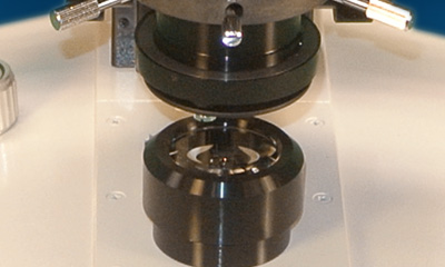
• Stage: Mechanical X-Y stage with attached finger assembly and drop down post, attached stage is 120mm x 140mm rectangular mechanical stage with movements of 75mm (X) x 40mm (Y) and is graduated with Vernier readings to 0.1mm, equipped with low positioned drop down coaxial controls. (Factory Installed)
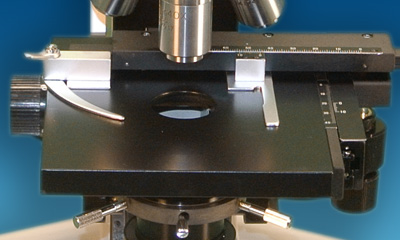
• Focusing Adjustment: Coaxial coarse and fine focusing controls with graduation reading 2 microns power division. Incorporates tension adjustment and safety Auto-focus stage stop lever

• Nosepiece: Reverse Facing Quadruple
• MA289/05: Blue filter, clear, 29.8mm diameter
• MA676: Attachable clinical specimen holder for MA596R and MA596L
• MA595R: Attached Graduated mechanical stage, 30mm X 70mm movement with drop-down coaxial controls (right) for the ML series ( left hand available upon request)
• MA326: Bulb 6V 30W Halogen ( Factory Installed)
• MA327: Fuse 3Amp (Factory Installed)
• MA700: Immersion Oil. 15cc
• MA701: Dust Cover for ML5000
• Manual: Meiji Techno America ML5000 Manual
• Warranty: Limited Lifetime Warranty Card
Product Information:
• Manufactured: 100% MADE IN JAPAN
• Warranty: LIMITED LIFETIME WARRANTY
• Customer Support: 1-(800) 832-0060 (U.S.A Based)
Package Information:
• Weight: 21.0 lbs (9.5 kg)
• Box Size: 22 x 16 x 23 inches
• Footprint: 254mm (D) x 203mm (W)
• Binocular head type: 255 mm(D) x 395 mm (H) x 195mm(W) 21.0 lbs. (9.5kg)
• Trinocular head type: 255 mm(D) x 440 mm (H) x 195mm(W) 22.0 lbs. (10.0kg)
|
Model
|
Head
|
Illumination
|
Eyepieces
|
Condensers
|
Objectives
|
Stage
|
|
ML5870 |
Binocular
|
Koehler
12V 50W halogen |
KWH10x FN20
|
Zernike condenser, NA 1.25 with GIF 546nm, 29.8mm Ø
|
BF Semi Plan Phase Contrast 10X, S20X, S40X, S100X oil
|
Flat-Top
|
|
ML5970 |
Trinocular |
VIEWING HEADS
Available with standard binocular heads or trinocular heads for photomicrography, 30° inclined eyetubes with adjustable diopter settings and interpupillary distance, 360° rotatable.
EYEPIECES
Paired DIN KHW10x with 20mm field of view.
OBJECTIVE CHANGER
Smooth-operating, ball bearing mounted, quadruple nosepiece.
OBJECTIVES
S. Flat Achromats, SM Planachromats, Planachromats, and Phase Contrast.
STAGE
Flat-top stage: 171mm x 140mm surface with 80mm (X) by 52mm (Y) movement, graduated with vernier readings to 0.1 mm, detachable specimen guide, drop-down right of left-handed controls.
CONDENSERS
Standard models have either an Achromatic NA 1.25 condenser with iris diaphragm and filter tray for brightfield work, simple NA 1.25 slider phase condenser, or an NA 1.25 Zernike phase condenser with filter tray and independently centerable annuli for phase contrast. Cardioid-type NA 1.25 darkfield condenser for high power (100x objective only) darkfield work is optional.
ILLUMINATOR
Variable-intensity Koehler illumination for apertures up to 1.25. Available in 6V 30W (built-in) or 12V 50W (attached lamp house) models.
The ML5870 features include:
- Transmitted light Binocular microscope with DIN standard eyepieces with 20mm field of view, 22mm eye relief
- Binocular head is inclined at 30° degrees and rotatable 360° degrees
- Dual graduated diopter eyetubes with interpupillary adjustments of 52mm to 72mm
- Trinocular models has a sliding beam splitter that directs 100% of the light to the eyepieces or 80% to the camera and 20% to the eyepieces
- Reverse facing, quintuple ball bearing smooth operating, revolving nosepiece
- SM Plan Objectives, 160mm Tube length, Non-Infinity objectives
- Flat-Top Mechanical stage with 171mm and 140mm rectangular stage with movement of 78mm (X) x 52mm (Y) with drop down
- Achromatic N.A. 1.25 condenser with iris diaphragm and filter carrier
- Coaxial coarse and fine focus adjustment with graduation, reading 2 microns per division with tension adjustment and safety auto focus stop
- Built-in Koehler illuminator with 6V 30W/12V 50W halogen lamp & variable intensity control
Brightfield:
The simplest of all the optical microscopy illumination techniques. Sample illumination is transmitted (i.e., illuminated from below and observed from above) white light and contrast in the sample is caused by absorbance of some of the transmitted light in dense areas of the sample. Brightfield microscopy is the simplest of a range of techniques used for illumination of samples in light microscopes and its simplicity makes it a popular technique. The typical appearance of a brightfield microscopy image is a dark sample on a bright background.
Phase Contrast:
An optical microscopy technique that converts phase shifts in light passing through a transparent specimen to brightness changes in the image. Phase shifts themselves are invisible, but become visible when shown as brightness variations. When light waves travel through a medium other than vacuum, interaction with the medium causes the wave amplitude and phase to change in a manner dependent on properties of the medium. Changes in amplitude (brightness) arise from the scattering and absorption of light, which is often wavelength-dependent and may give rise to colors. Photographic equipment and the human eye are only sensitive to amplitude variations. Without special arrangements, phase changes are therefore invisible.
The ML5000 Series has a Koehler Halogen 6V 30W or A 12V 50W illumination with a reverse facing quintuple ball bearing nosepiece and auto voltage sensing power supply. High resolution DIN Standard SM Plan objectives offer crisp, clear, high contrast images and are offered as part of the standard package. A built in integrated Koehler illuminator with a field iris and 6V 30W/12V 50W Halogen lamp and variable intensity control is provided standard equipment to complement the SM PLAN optics. The low voltage, high intensity, pre-centered halogen lamp provides high level illumination can be adjusted to maximize the evenness of the bulb illumination and is readily accessible for a quick bulb replacement. Flat-Top Mechanical stage with 171mm and 140mm rectangular stage with movement of 80mm (X) x 52mm (Y) with drop down Ergonomically placed low positioned coaxial controls and a detachable specimen guide.


